Phylum Rotifera
http://en.wikipedia.org/wiki/rotifer
General:
Rotifers are microscopic, multicellular organisms that are commonly found in marine and freshwater environments .The protozoans seem to be the most diverse group on the bog with the rotifers a close second. Videos shown below showcase the anatomy of a typical bdelloid rotifer.

A ciliated bi-lobed disc (Corona) is attached to the body as shown in the first and second video. A long antenna is visible just below the corona toward the end of the second movie. It can be retracted inside the mouth (2nd video) when the rotifer is disturbed. Disc cilia move the unattached animal smoothly through the water. When the rotifer is attached to the substratum the cilia create water currents (Videos 1 and 2) that move food ( Bacteria, detritus, small protozoans, etc.) into the mouth. Food then passes down a short passageway (Visible) into a constantly beating structure called the mastax (Visible). The mastax is made up of muscles and hard jaws (Trophi) that grasp and grind up the food before it is passed through a short oesophagus into the large brownish stomach (Visible). Here food is digested and the indigestible remains pass into a short intestine (Visible) and then to the outside through the anus.(Photographs of Trophi from a large number of rotifer species are available at http://rotifer.acnatsci.org/rotifer.php/trophi. The body is covered with a cuticle that is secreted by the hypodermis. The cuticle is ringed with thin annuli allowing the animal to extend or shorten the body in telescopic fashion. A short Foot (Visible in Video 2) extends from the posterior part of the body and gives rise to four terminal toes (Visible). The toes secrete a mucus-like material that holds the rotifer in place while they feed. Two small red eyes are visible in the first movie just below the ciliated corona. A single, relatively large ,squarish ,bladder (Second video) is visible just below the short intestine. It contracts periodically, passing water taken into the body osmotically,to the outside.
http://vimeo.com/82147756 (Zottoli) (Philodina)
http://vimeo.com/81766056 (Zottoli) (Philodina)
Description of HVNC Rotifers:
1., Bryceella(Proalidae)
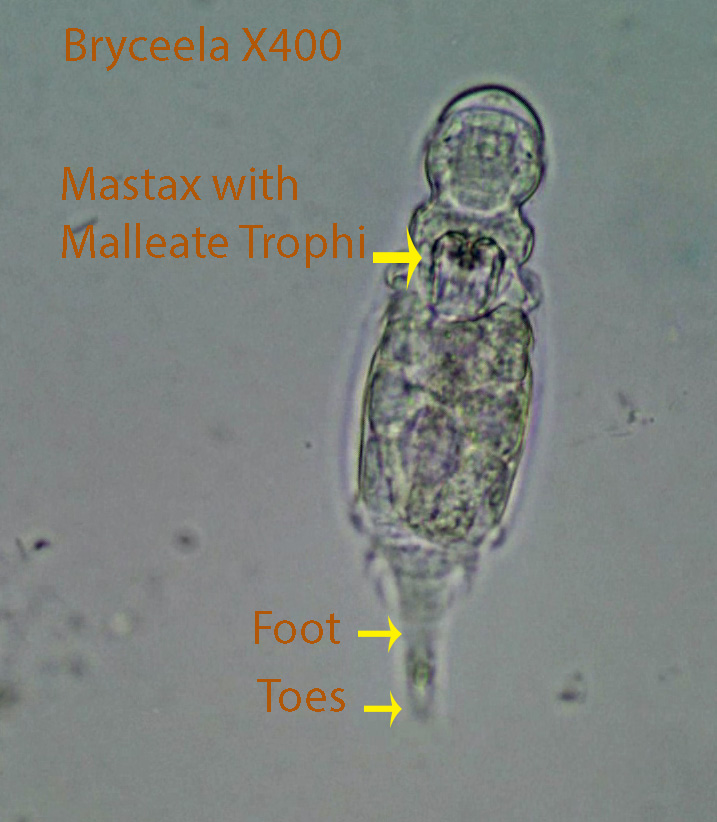
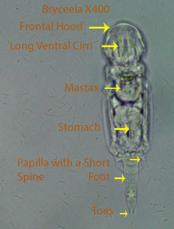
Long ventral cirri that point posteriorly are found on the anterior ventral portion of the head. An anterior dorsal rostrum (Frontal Hood) extends from the front part of the head downward.The long ventral cirri and the relatively short ventral cilia that follow them, dislodge food particles from the substratum. The actively moving mastax with its malleate trophi was observed extending out of the mouth and grabbing food particles in front of it. The mastax passes food through a short oesophagus into the stomach where digestion takes place. One small papilla with a small spine (Stylus)is present laterally on each side of the anterior portion of the head. The styli are small in Type 2. The outside covering (Lorica) surrounding the rotifer extends posteriorly over the dorsal surface for a short distance. Below this extension the foot and two toes are visible. The toes are able to temporarily attach to the substratum, allowing the animal to pivot around and move quickly to another spot. A short spine is present dorsally at the end of the foot. A long spine is present on the dorsal surface of type 2 only.
The animal moves erratically with quick spastic movements, feeding for a moment, and then moving to another place.
http://vimeo.com/81895735 (Zottoli) (Bryceella)
http://vimeo.com/84494297 (Zottoli) (Bryceella)
http://vimeo.com/84494298 (Zottoli) (Bryceella)
http://vimeo.com/85207759 (Zottoli) (Bryceella Type 2)
http://vimeo.com/85207758 (Zottoli) (Bryceella Type 2)
2. Cephalodella Type 1 (Notommatidae)

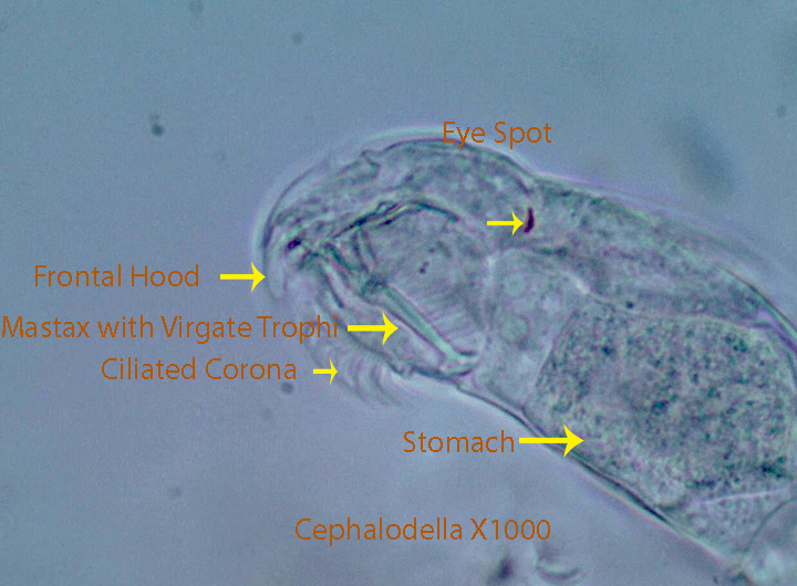
An anterior rostrum (Frontal Hood) extends downward from the front part of the head. Cilia on the ventral part of the head seem to dislodge food particles (Detritus, Bacteria, small protozoans, etc.) from the substratum. The mastax with its virgate trophi, appears to draw food particles inward and passes them through a short oesophagus to the stomach where digestion takes place. Digested material passes into the small intestine and then out through the anus. They often feed on the surface of Sphagnum leaves or on clumps of debris containing bacteria and protozoans. In one instance the stomach turned green as they fed along the edge of a Sphagnum leaf.
http://vimeo.com/84548117 (Zottoli) (Cephalodella X1000. Note the rostrum, ventral cilia, virgate trophi and red eye spots)
http://vimeo.com/82149817 (Zottoli) (Cephalodella Type 1) X400
http://vimeo.com/84548115 (Zottoli) (Cephalodella Type 1 ) X400
3. Cephalodella , Type 2 X400
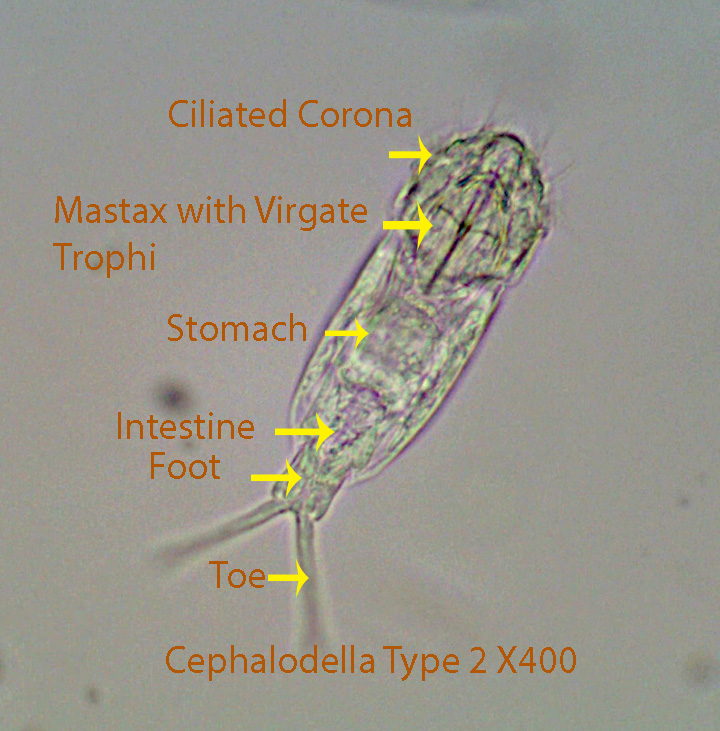
Cephalodella Type 2 , about 90 microns long, has a ciliated corona that circulates water as shown in the second and last movies. Small prey tend to fall towards the center of the corona within easy reach of the constantly active. apical mastax and its virgate trophi. The trophi are thrust out of the mouth quickly and brought back into the mouth, presumably with small prey. The whole process is too fast to clearly see what is happening. The ciliated stomach of the rotifer in the third movie appears to contain bacteria only. During the feeding process the rotifer usually attaches its two toes to the substratum and extends its body outward into the water column. In one instance (Video 3) , the toes are attached and the head is propped up on a mound of debris by stationary long, ventral coronal cilia, during a feeding bout. The mastax pushes food through the short oesophagus into the stomach (Visible) where digestion takes place. Undigested food passes into the intestine where more digestion may take place before it is forced to the outside through the anal opening. A clear, contractile bladder lies behind the intestine. The next to last last video, shows a lateral view of the virgate trophi, that are typical of Cephalodella.
https://vimeo.com/85348399 Cephalodella Type 2 X400
https://vimeo.com/85348401 Cephalodella Type 2 X400
4. Collotheca (Collothecidae)


Collotheca is a large rotifer, about 550 microns long. The anterior corona is made up of several lobes with long setae that extend outward into the water column. The setae contract slowly suggesting that they are modified cilia. The coronal lobes and setae often fold inward trapping any prey that find their way into the funnel-shaped space below the lobes. When this happens, prey (Protozoans and other rotifers for example) are pushed downward and passed through the mouth into a large chamber (Proventriculus). The Mastax, with its uncinate trophi,located at the bottom of the proventriculus, moves food into the ciliated stomach where digestion takes place. Round,usually darkened grape-like gastric glands lie around the stomach. Empty rotifer loricas in the proventriculus of a number of specimens suggest that some pre-digestion may occur here. I observed a protozoan swimming into the proventicular chamber of a fully extended specimen.It was eventually grabbed by the mastax and passed directly into the stomach while the rotifer remained in an extended position. The rotifer is usually attached to the substratum and can move up and down through contraction and extension of its accordion-like foot.
http://vimeo.com/81763496 (Zottoli) (Collotheca X400)
http://vimeo.com/81763497 (Zottoli) (Collotheca X400 showing the mastax in action)
http://vimeo.com/55044626 (Zottoli) (Collotheca X400 showing the contractile tail)
http://vimeo.com/81763495 (Zottoli) (Collotheca X400)
5.Collurella (Lepadellidae)

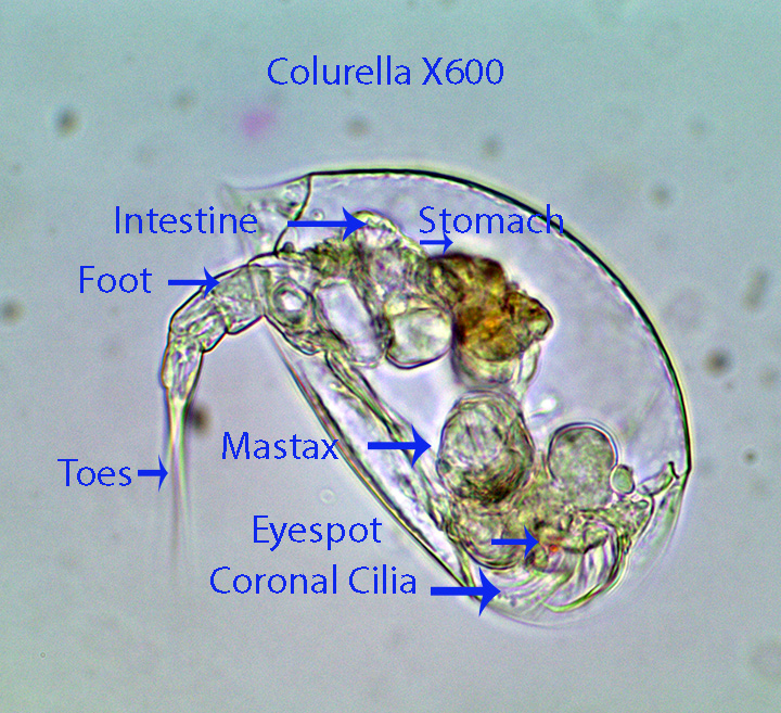
The rigid lorica around the rotifer is composed of two lateral plates that are strongly compressed. The rotifer, about 120 microns long, has a ciliated corona that is responsible for moving the animal forward. The toes sometimes are attached to the substratum and the animal then bends downward touching the bottom with its cilia. The movement of coronal cilia dislodge food particles that are then directed to the mouth. A frontal hood extends downward in front of the face. It is positioned in such way that as the corona is withdrawn into the lorica, it may prevent food from escaping to the sides. Contraction of the foot in concert with the positioning of the toes allows the animal to turn and twist. The active mastax with its malleate trophi passes food through a short oesophagus into the stomach. Small green flagellates as well as bacteria have been seen inside the stomach. The ciliated corona can be completely withdrawn into the lorica.
http://vimeo.com/84637887 (Zottoli) (Colurella)
http://vimeo.com/84637888 (Zottoli) (Colurella)
6. Conochilus (Conochilidae)

Conocoelis is an elongated rotifer with an anterior , circular, ciliated corona. A mid ventral portion of the corona lacks cilia. The annulated foot is unsegmented. The intestine is u-shaped and opens towards the anterior end (The wider arrow associated with the intestine label is pointing at the ascending intestinal tube). This specimen was probably part of a large colony of like individuals.
https://vimeo.com/172036377 Conochilus X400
https://vimeo.com/172038614 Conochilus X400
https://vimeo.com/172036348 Conochilus X400
7. Dicranophorus (Diocranophoridae)

Dicranophorus, about 325 microns long, has a frontal hood that drapes over the anterior end, partly covering the ciliated ventral corona. The strong coronal cilia draw the animal smoothly forward. The long, wide toes allow the rotifer to quickly turn in any direction. During a turning maneuver, the toes first push against the substratum and thus allow the rotifer to pivot in the desired direction. The mastax has unique forcipate/virgate trophi with slender, pointed anterior jaws designed to penetrate and hold prey. Although I was unable to see feeding,the structure of the mastax suggests that the rotifer is most likely a predator. Two eye spots are visible on most specimens. Prey are passed by the mastax into a large stomach where digestion occurs. The indigestible remains are pushed into the intestine and then out of the anus. A clear, rounded Bladder is visible at the end of the body not far from the posterior part of the intestine. A relatively narrow foot is followed by two toes.
http://vimeo.com/85620118 (Zottoli) (Dicranophorus X400 Anatomy Movement)
http://vimeo.com/82219617 (Zottoli) (Dicranophorus X100. Movement)
http://vimeo.com/82219619 (Zottoli) (Dicranophorus X400 Anatomy)
http://vimeo.com/82219620 (Zottoli) (Dicranophorus X400 Anatomy)
http://vimeo.com/82219613 (Zottoli) (Dicranophorus X400 Anatomy)
http://vimeo.com/84782567 (Zottoli) (Dicranophorus X400 Anatomy)
http://vimeo.com/84782568 (Zottoli) (Dicranophorus X400 Virgate Trophi)
8. Dissotrocha (Philodinidae)
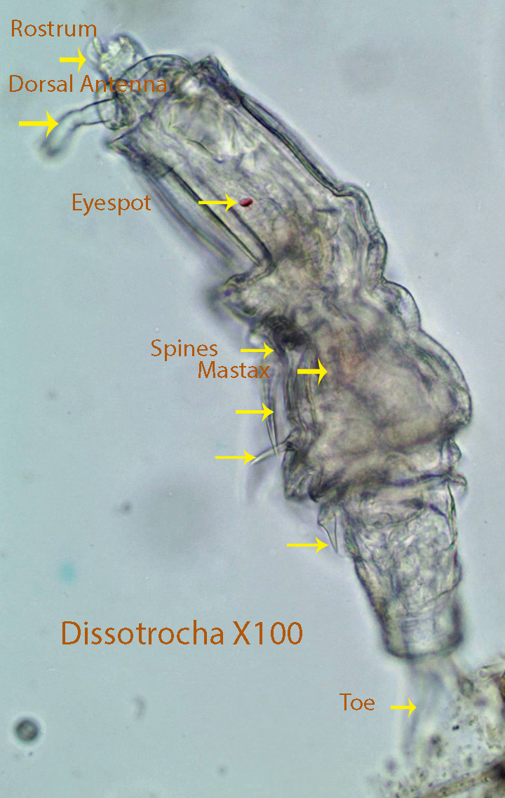
Dissotrocha, about 500 microns long, has two anterior ciliated discs (Trochal Discs). They can be seen about mid-way through the last video. In all the other videos they are retracted internally. They look like two rounded muscular spheres just below the anterior end. The outside covering (Integument) is not flexible and is characterized by stout projecting spines. A ciliated finger-like structure (Rostrum) projects anteriorly from the dorsal part of the head. There is a smaller finger-like projection (Dorsal Antenna) that extends laterally below the rostrum. Both are sensory projections that provide information about the environment around them.
http://vimeo.com/82690274 (Zottoli) (Dissotrocha)
http://vimeo.com/84803918 (Zottoli) (Dissotrocha X400 Body Spines)
http://vimeo.com/84804644 (Zottoli) (Dissotrocha X 400 Trochal Discs)
9.Dorystoma (Notommatidae)

Dorystoma, about 230 microns long, is able to move rapidly in a straight line propelled by the anterior coronal cilia. Movement appears to be random until a clump of debris or a Sphagnum leaf is reached. It feeds and then moves randomly until it encounters another source of food. It appears to consume detritus bacteria, small protozoans, etc. The rotifer can also turn by moving the posterior third of its body in the direction that it chooses. Afterwards, the animal straightens out and moves along. Sometimes the animal plants its toes against the substratum, and then bends its body in the direction that it chooses and moves forward. When the rotifer feeds, it centers its anterior end over clumps of debris and the coronal cilia push food towards the ventral mastax which forces food into the stomach. The genus is characterized by a dorsal spine on the distal end of the body as well as a pair of lateral outgrowths each with a relatively short spine.
http://vimeo.com/82691967 (Zottoli) (Dorystoma)
http://vimeo.com/82690952 (Zottoli) (Dorystoma)
http://vimeo.com/82690953 (Zottoli) (Dorystoma)
http://vimeo.com/85628129 (Zottoli) (Dorystoma X400 Movement, Anatomy)
http://vimeo.com/85628131 (Zottoli) (Dorystoma X400 Anatomy,Movement,Feeding)
http://vimeo.com/85628694 (Zottoli) (Dorystoma X400 Movement, Anatomy)
http://vimeo.com/85628693 (Zottoli) (Dorystoma X400 Movement,Anatomy)
10. Filinia spp. (Trochosphaeridae)
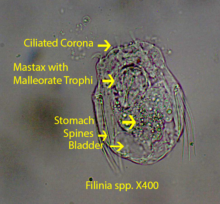
Filinia, about 140 microns long, is characterized by movable anterior spines that push against the substratum causing the rotifer to “hop” from place to place. There are two anterior spines on each side of the head and one rigid spine on each side near the posterior end. The animal lacks a foot and toes. The coronal cilia create a circular water flow from the sides of the lobes to above the head and back down again. Detritus, bacteria and small protozoans drop downward towards the mouth. The mastax with its malleoramate trophi seem to grasp food (Detritus, bacteria, small protozoans, etc.)and push it into the stomach.
http://vimeo.com/82694877 (Zottoli) (Filinia X400 Anatomy)
http://vimeo.com/82737274 (Zottoli) (Filinia X100 Movement)
http://vimeo.com/82737276 (Zottoli) (Filinia X400 Egg)
http://vimeo.com/82737275 (Zottoli) (Filinia X400 Mastax with Malleoreate Trophi pushing into Stomach)
11. Habrotrocha (Habrotrochidae)

Habrotrocha, about 300 microns long, is often found in a mucus burrow secreted in layers as shown in the last movie. This rotifer is characterized by its lack of a stomach lumen. Instead the stomach is formed of individual, round cells that most likely consume food much like an amoeba. Small, ciliated ,trochal discs create a circular water flow from the sides of the lobes to above the head and back down again. Detritus, bacteria and small protozoans drop downward towards the mouth and then into a ciliated oesophagus that leads into the beating mastax. The mastax has ramate trophi that grind up the food and push it into the stomach where digestion takes place. There are two red-eye spots just above the mastax, that can be seen in several of the videos. They are capable of moving forward smoothly pulled by the action of the trochal cilia. A second type of movement characteristic of many species of rotifers involves first attaching their toes to the substratum and then extending their body in the direction of movement. During the next phase, they attach their head and pull the posterior end to just behind the head moving somewhat like an inch worm. The process is repeated again and again as the animal moves forward. The trochal discs are retracted when they move in this way.
http://vimeo.com/82738289 (Zottoli) (Habrotrocha X400 Anatomy)
http://vimeo.com/82739512 (Zottoli) (Habrotrocha X100 Movement)
http://vimeo.com/82738291 (Zottoli) (Habrotrocha X400 Feeding)
http://vimeo.com/82739513 (Zottoli) (Habrotrocha X400 Mucus Burrow)
12.Keratella (Brachionidae)

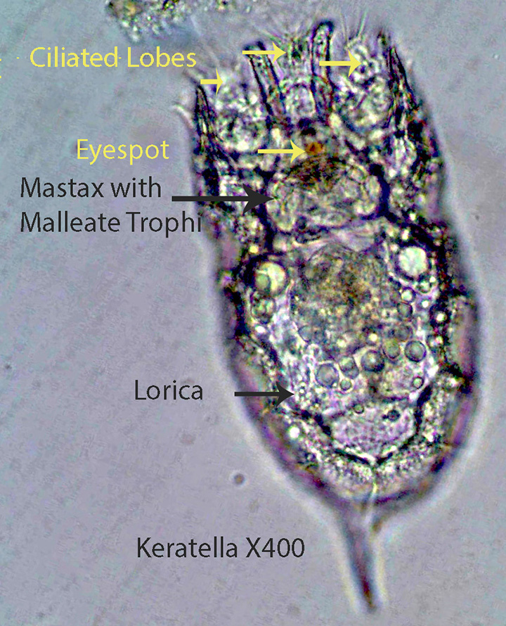
Keratella, about 160 microns long, is characterized by its elongate anterior spines and complete lack of a foot and toes. The rotifer is covered by a distinct rigid lorica consisting of a dorsal and a ventral plate. The rotifer moves somewhat erratically propelled by the coronal cilia. As they move forward they rotate around the anterior-posterior axis. The ciliary lobes create a circular current of water that passes from the anterior end, posteriorly along the sides of the animal and then back anteriorly in a circular loop. Food (Detritus, bacteria, small protozoans, etc.)tends to drop out of the loop above the head and is actively grabbed by the malleate trophi in the mastax. The mastax macerates the food and forces it into the stomach through a short oesophagus. A round, clear bladder is present near the posterior end.
http://vimeo.com/82215792 (Zottoli) (Keratella X100 Movement)
http://vimeo.com/82215793 (Zottoli) (Keratella X400 Anatomy and Feeding)
http://vimeo.com/82215794 (Zottoli) (Keratella X400 Anatomy and Feeding)
http://vimeo.com/81896743 (Zottoli) (Keratella X400 with Egg)
13. Lecane (Lecanidae)

Lecane, about 120 microns long, is covered by a thick, inflexible lorica with a lateral groove that connects the dorsal and ventral plates. The mastax with malleate trophi, is active and applied to the substratum during feeding. Generally the toe(s) attach to the substratum,the body bends downward, and the ventral mastax using the trophi, pulls food (Detritus, bacteria, small protozoans, etc.) inside and passes it into the stomach. This process is visible in the last movie. Lecane has a short foot and either a single toe (Type A) or two toes (Type B). The two toes of type B rotifers may be fused at the tip or completely separate. The coronal cilia alone can move the animal, however the toes can push against the substratum and turn the animal in a different direction.
http://vimeo.com/84962869 (Zottoli) (Lecane X400 Type A)
http://vimeo.com/82238755 (Zottoli) (Lecane X400 Anatomy Type B)
http://vimeo.com/82238756 (Zottoli) (Lecane X400 Anatomy Type B)
http://vimeo.com/84962870 (Zottoli) (Lecane X400 Feeding)
14. Lepadella (Lepadellidae)


Lepadella, about 120 microns long, is dorso-ventrally flattened . The dorsal and ventral plates of the lorica are completely fused. The rotifer lacks a frontal hood like that found on Colurella. The ciliated corona can be extended or retracted through an anterior opening in the lorica. The foot extends to the outside through a ventral notch in the lorica. During the feeding process, the rotifer contacts the surface , attaches the tips of the toes, and bends at about a 30 degree angle with the coronal cilia near the substratum. The coronal cilia seem to dislodge material from the substratum. The food is carried by cilia for a short distance and grabbed by the ventral malleate trophi (Part of the Mastax) and in concert with contractions of Mastax muscles, food is pushed through a short oesophagus into the stomach. The coronal cilia pull the animal forward. As they move forward they also rotate around their anterior-posterior axis. The rotifer also uses its toes to set up a pivot point that allows them to move their body and change direction. Four types of this genus can be separated by the structure of their lorica . The lorica of Type 1 is relatively smooth while the lorica of Type 2 has a pronounced mid dorsal ridge extending the length of the lorica. The lorica of Type 3 has obvious dorsal ridges while that of Type 4 has dorsal ridges but also posterior, pointed, lateral edges .
http://vimeo.com/82241138 (Zottoli) (Lepadella X400 Type 1 Anatomy)
http://vimeo.com/82242286 (Zottoli) (Lepadella X400 Type 1 )
http://vimeo.com/82241140 (Zottoli) (Lepadella X100 Type 1 Movement)
http://vimeo.com/82238754 (Zottoli) (Lepadella X100 Type 2 Movement )
http://vimeo.com/82242285 (Zottoli) (Lepadella X400 Type 3 )
http://vimeo.com/82242283 (Zottoli) (Lepadella X400 Type 4 )
http://vimeo.com/82147760 (Zottoli) (Lepadella X400 feeding on the edge of a Sphagnum leaf)
15. Microcodon (Microcodinidae)
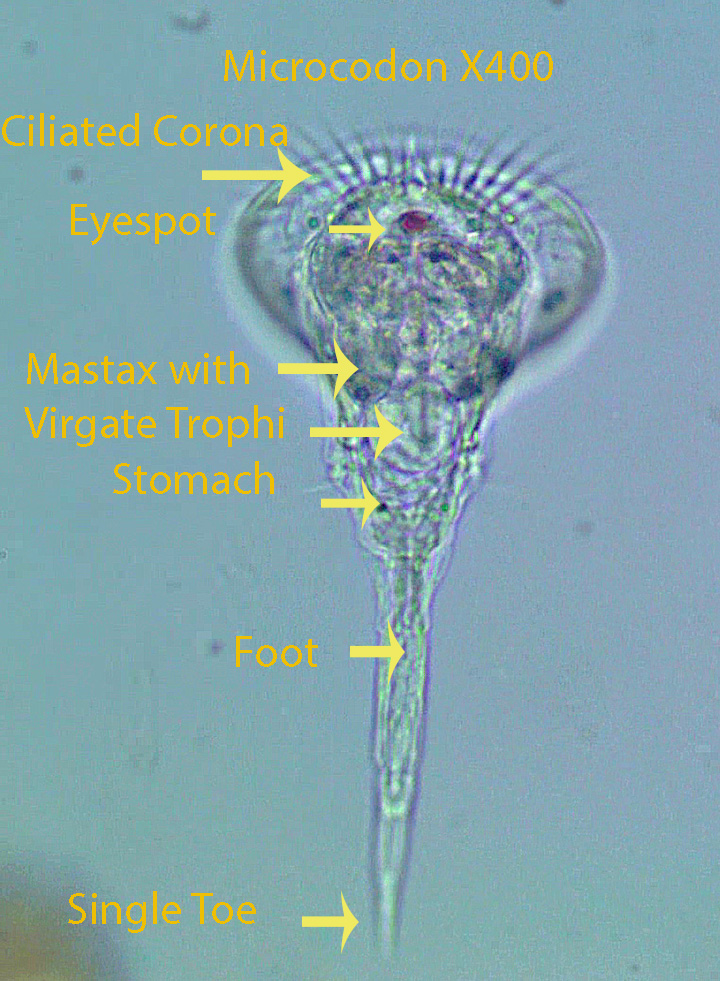
Microcodon, about 470 microns long, has a wide, flat, circular, ciliated corona. One red eye-spot is located just below the corona. The mastax with virgate trophi is situated slightly below the corona and connects through a short oesophagus to the wider stomach. A pair of small lateral papillae each with a short spine are visible towards the end of the tapered body. A clear rounded bladder lies below the papillae. The body ends a short distance from the bladder. A long foot with a spear-like toe, is attached to the end of the body. The animal can move in a straight line powered by the coronal cilia or it can attach its single toe to an object and move to the right or left or in a circle. Feeding was not observed.
http://vimeo.com/85002723 (Zottoli) (Microcodon X100 Movement)
http://vimeo.com/85002722 (Zottoli) (Microcodon X400 Anatomy)
http://vimeo.com/85002720 (Zottoli) (Microcodon X400 Anatomy)
16. Monommata (Notommatidae)

Monommata, about 350 microns long, is characterized by its long toes (Longer than the body) and short foot. A dark eye spot lies below the oblique corona and the mastax with its virgate trophi is located near the eye spot. The ventral mastax with its trophi can be protruded from the mouth and pull food (Detritus, bacteria, small protozoans, etc.) into the mouth and push it into the stomach. The rotifer can move forward in a straight line powered by the coronal cilia. The animal can change direction by placing its toes on the substratum, bending its body and moving on a new path.
http://vimeo.com/55235338 (Zottoli)( Monmmata X100 Movement)
http://vimeo.com/82247048 (Zottoli) (Monommata X400 Anatomy)
http://vimeo.com/82248964 (Zottoli) (Monommata X400 Compressed, Anatomy)
17. Mytilina (Mytilinidae)

Mytilina , about 350 microns long, is characterized by its firm ,thick lorica ,made up of two lateral plates and one ventral plate. Spines extend from the anterior corners of the lateral plates. The mastax with its malleate trophi are visible below the ciliated corona. One red eyespot is located just above the mastax. The mastax pushes against the large brown stomach. The intestine lies just below the stomach. A short foot and two relatively long toes can be seen towards the end of the video.
http://vimeo.com/82248965 (Zottoli) (Mytilina)
18. Notommata (Notommatidae)

Notommata, about 350 microns long, has a ventral mastax with virgate trophi that lies just behind the ventral coronal cilia. There is a distinct separation between the head and the body. A pair of small, lateral, ciliated lobes is located just behind the rounded anterior part of the head. They can be seen at about the middle part of the third video. The vitellarium, visible in all three videos,is rounded. The body is flexible (Illoricate). The foot and toes are small and dorso-ventrally flattened. There are no frontal eye-spots or spines on the body. A relatively large dorsal papilla (Fleshy Extension) is present at the end of the body. It can best be seen in the third video.
http://vimeo.com/82308162 (Zottoli) (Notommata X400 Anatomy)
http://vimeo.com/82310445 (Zottoli) (Notommata X400 Anatomy)
http://vimeo.com/85733829 (Zottoli) (Notommata X400 Anatomy,Dorsal Papilla, Small Anterior lateral Lobes)
19. Philodina (Philodinidae)
Philodina, about 450 microns long, has two ciliated coronal (Trochal) lobes that extend into the water column while feeding. The first video shows the circular current powered by the trochal cilia. Food tends to fall out of the circular current over the mouth where it is moved by cilia to the mastax fitted with hard rami that macerate and push food through the oesophagus into the stomach for processing. Food then passes into the intestine and undigested material is pushed to the outside through the anal opening. When trochal cilia are extended and the toes are not attached, the cilia draw the animal forward in a straight line. When the toes are attached to the substratum and the ciliated trochal discs are extended, the cilia circulate the surrounding water bringing food to the mouth. A second type of movement characteristic of many species of rotifers involves first attaching the toes to the substratum and then extending their body in the direction of movement. During the next phase, they attach their head and pull the posterior end to just behind the head moving somewhat like an inch worm. The process is repeated again and again as the animal moves forward. During this process the coronal discs are retracted.
http://vimeo.com/85104701 (Zottoli) (Philodina X400 Anatomy Movie1)
http://vimeo.com/85102516 (Zottoli) (Philodina X400 Anatomy)
http://vimeo.com/85104702 (Zottoli) (Philodina X400 Feeding Currents, Mastax)
http://vimeo.com/82147756 (Zottoli) (Philodina X400 Feeding)
http://vimeo.com/81766056 (Zottoli) (Philodina X400 Feeding)
http://vimeo.com/85104700 (Zottoli) (Philodina X400 Bladder Contraction)
http://vimeo.com/85102515 (Zottoli) (Philodina X400 Trochal Cilia Missing)
20. Platyias (Brachionidae)

The animal, is about 160 microns long. Platyias has a mastax with malleate trophi as well as a short foot and two toes. The spiny lorica as shown in the video is a characteristic of the genus. This rotifer is rare at the HVNC bog.
http://vimeo.com/82245983 (Zottoli) (Platyias)
21.Proalinopsis (Proalidae)


Proalinopsis, about 500 microns long, has a mastax with malleorate trophi. When the rotifer feeds, it places its ciliated corona slightly above a clump of debris.The coronal cilia sweep the surface shaking loose small organisms (Bacteria and small protozoans, etc.) as well as detritus. This material is guided by cilia into the mastax where it is somewhat macerated and then passed into the stomach through a long ciliated oesophagus (Visible). Digestion takes place in the stomach and the prominent gastric glands that empty into the stomach most likely secrete digestive enzymes. Indigestible matter is passed into the intestine and eventually to the outside through the anal opening. A foot with two toes extends beyond the body. Also a papilla bearing a relatively long spine protrudes from the dorsal surface at the end of the body just before the beginning of the foot. In some specimens there are two small lateral papillae each bearing a spine (Visible in the second photograph) at about the middle of the body.
http://vimeo.com/82314819 (Zottoli) (Proalinopsis X400 Anatomy)
http://vimeo.com/82314820 (Zottoli) (Proalinopsis X 400 Anatomy)
http://vimeo.com/85202745 (Zottoli) (Prolinopsis X400 Excellent Anatomy)
http://vimeo.com/85202746 (Zottoli) (Prolinopsis X400 Mastax and Coronal Cilia)
http://vimeo.com/85202744 (Zottoli) (Prolinopsis X400 Feeding)
22.Pyturga (Fosicularidae)
Pyturga, about 424 microns long, is a sessile, filter feeding rotifer with an elongate body, that lives in a gelatinous tube. The tube is attached to a Sphagnum leaf in the movie below. The circular, ciliated corona creates water currents that move food to the center of corona where it is passed to the active mastax equipped with malleorate trophi and pushed into the stomach.
https://vimeo.com/116272310 Pyturga X400
23.Rotaria (Philodinidae)
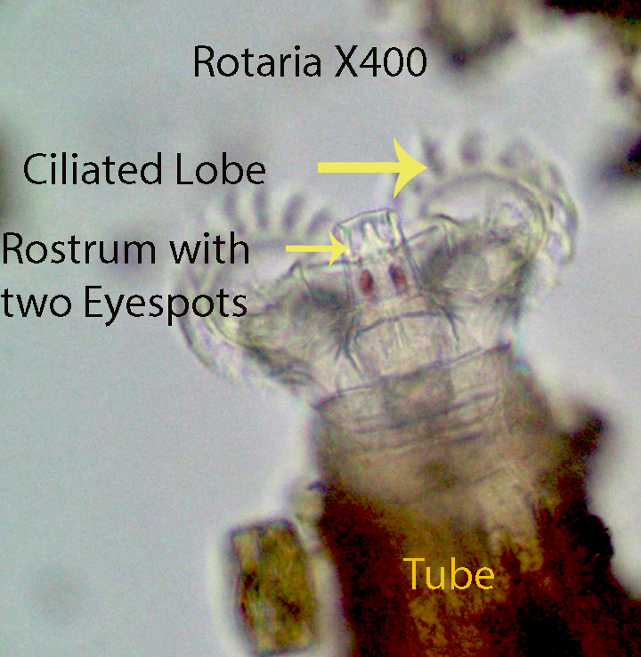
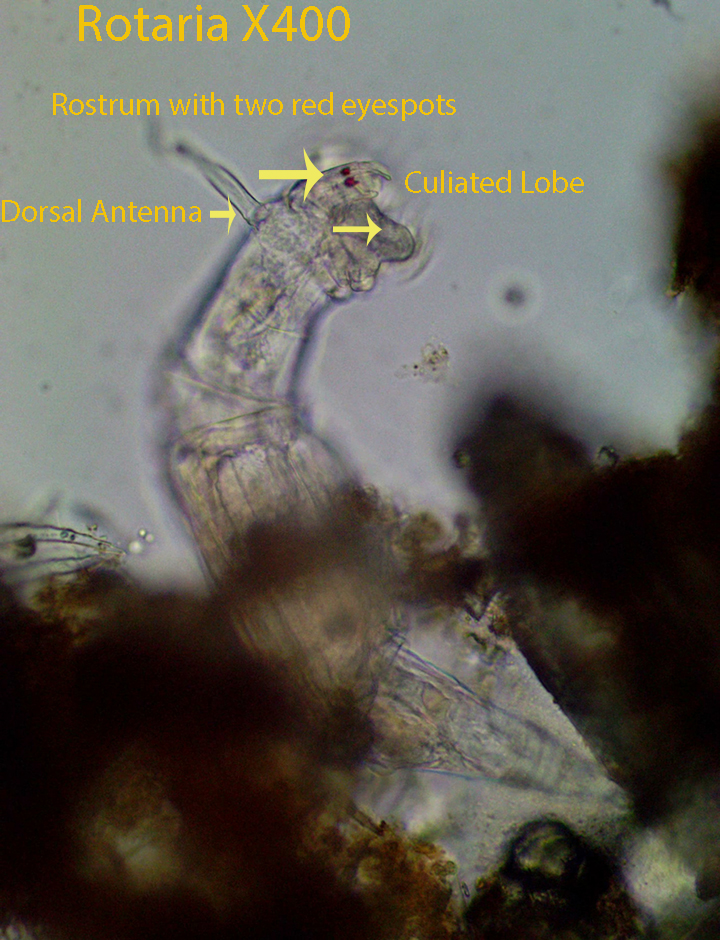
Rotaria, about 450 microns long, extends two ciliated coronal (Trochal Discs) lobes out into the water column while feeding. A rostrum that lies between the trochal discs has two distinctive red-eye spots (Shown best in the third video). The first and second movie show the circular water currents powered by the trochal cilia. Food tends to fall out of the circular current over the mouth where it is moved by cilia to the mastax. The mastax with its hard rami, macerate and push food into the stomach for processing. Food then passes into the intestine and undigested material is pushed to the outside through the anal opening. When the trochal cilia are extended and the toes are not attached, the cilia draw the animal forward in a straight line. When the toes are attached to the substratum and the trochal discs are extended, the cilia circulate the surrounding water and drive food toward the mouth. A second type of movement characteristic of many species of rotifers involves first attaching their toes to the substratum and then extending their body in the direction of movement. During the next phase, they attach their head and pull the posterior end to just behind the head moving somewhat like an inch worm. The process is repeated again and again as the animal moves quickly forward. During this process the coronal discs are retracted. Like other members of the family Philodinidae there are three toes, one dorsal and two ventral.
http://vimeo.com/82322838 (Zottoli) (Rotaria X400 Feeding)
http://vimeo.com/85262927 (Zottoli) (Rotaria X400 Feeding)
http://vimeo.com/85264929 (Zottoli) (Rotaria X400 Rostrum with Eye spots)
24. Rousseletia (Notommatidae)
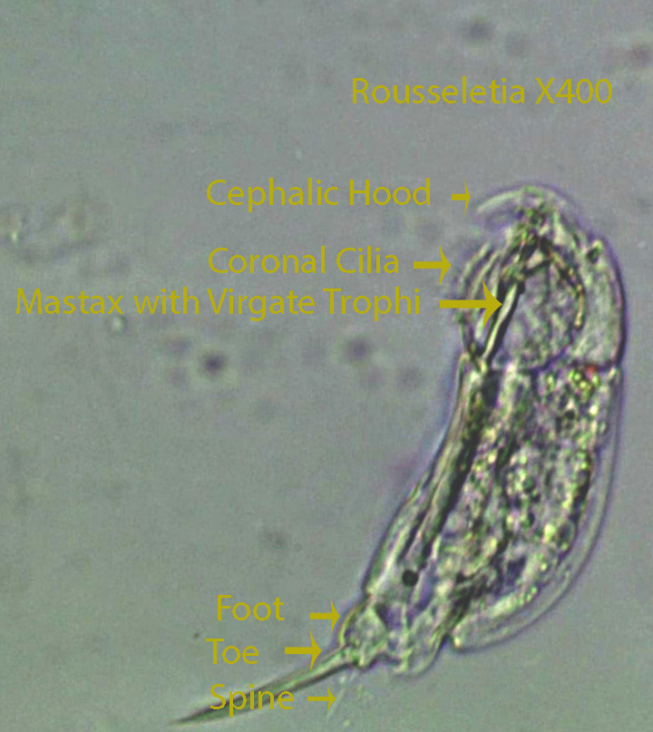
Rousseletia, about 120 microns long, is equipped with a mastax bearing virgate trophi and with a bean-shaped vitellarium. The toes (2) are shorter than the length of the body. A single dorsal spine is present at the end of the foot.
https://vimeo.com/85283651 Rousseletia X400
https://vimeo.com/82322840 Rousseletia X400
25. Squatinella Type 1 (Lepadellidae)

Squatinella, Type 1,about 160 microns long, is characterized by a wide dorsal shield that covers the ventral coronal cilia. Also two diverging spines arise just above the mastax and extend anteriorly. They can be seen in most of the videos. Two small red eyespots are located on the posterior lateral edges of the head. The mastax with its malleate trophi can be extended out of the mouth. The rotifer has a segmented foot and two, stiff, pointed toes.
Squatinella, Type 2 , about 160 microns long, has a smaller dorsal shield than Type 1 and lacks the anterior dorsal spines. Other than that it is similar anatomically to Type 1. In addition it has a long dorsal spine that is about the same length as the body. The sixth movie shows a Type 2 rotifer moving along the edge of a Sphagnum leaf. The long dorsal spine may make it more difficult for predators to handle and swallow the rotifer.It clearly extends the malleate trophi out of the mouth and appears to be scraping the leaf surface as it moves forward. There is a small dorsal spine projecting from the end of the foot. Movement is powered by the coronal cilia and aided by body contractions. When the toes are attached to the substratum they may act as a pivot point allowing the animal to turn and head off in a different direction.
http://vimeo.com/85302631 (Zottoli) (Squatinella X400 Type 1 Anatomy)
http://vimeo.com/85302629 (Zottoli) (Squatinella X400 Type 1 Anatomy)
http://vimeo.com/85290517 (Zottoli) (Squatinella Type 1 X400 Anatomy)
http://vimeo.com/85290515 (Zottoli) (Squatinella Type 1 X400 Anatomy)
http://vimeo.com/85294276 (Zottoli) (Squatinella Type 2 X400 Anatomy Movement)
http://vimeo.com/85294275 (Zottoli) (Squatinella Type 2 X400 Movement)
http://vimeo.com/85295561 (Zottoli) (Squatinella X400 Type 2 Anatomy)
http://vimeo.com/85303686 (Zottoli) (Squatinella X400 Type 2 Anatomy)
26. Synchaeta (Synchaetidae)

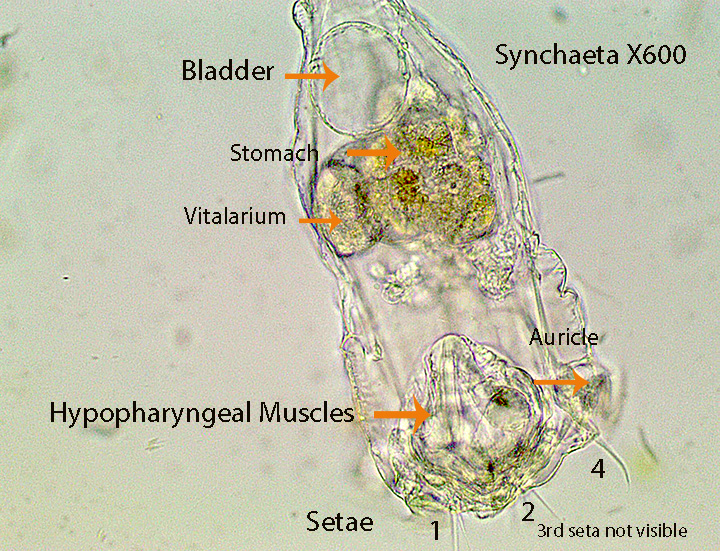
Synchaeta, about 400 microns long, has a mastax with virgate trophi, connected to large, visible, hypo-pharyngeal muscles. The lorica surrounding the rotifer is flexible (Illoricate). The ciliated corona is flanked on both sides by a group of lateral cilia. These most likely correspond to the “ear like lobes” listed as a characteristic of this genus. A pair of small, lateral, stiff setae are present at the point where the “head” ends and the “body” begins. The body tapers posteriorly joining a short foot and single toe. A large, brownish stomach lies behind the mastax and a small whitish vitellarium is situated next to the stomach in the anterior part of the body.
http://vimeo.com/85559867 (Zottoli) (Synchaeta X400 Hypopharyngeal Muscles in Mastax, Anatomy)
http://vimeo.com/85561011 (Zottoli) (Synchaeta X400 Feeding)
http://vimeo.com/85561010 (Zottoli) (Synchaeta X400 Feeding, Anatomy, Movement ,Lateral Head Spines)
http://vimeo.com/85561009 (Zottoli) (Synchaeta X400 Movement Anatomy)
http://vimeo.com/85560749 (Zottoli) (Synchaeta X400 Anatomy Movement)
http://vimeo.com/85559869 (Zottoli) (Synchaeta X400 Anatomy)
27.Taphrocampa (Notommatidae)

Taphrocampa, about 120 microns long, possesses a simple ciliated corona and a flexible lorica. The rotifer has a mastax with virgate trophi. The dorsal side of the body has characteristic deep folds. The body ends with a short foot and two small toes.
http://vimeo.com/82645776 (Zottoli) (Taphrocampa)
http://vimeo.com/85354556 (Zottoli) (Taphrocampa)
http://vimeo.com/85354554 (Zottoli) (Taphrocampa)
28. Testudinella (Testudinellidae)
https://species.wikimedia.org/wiki/Testudinella
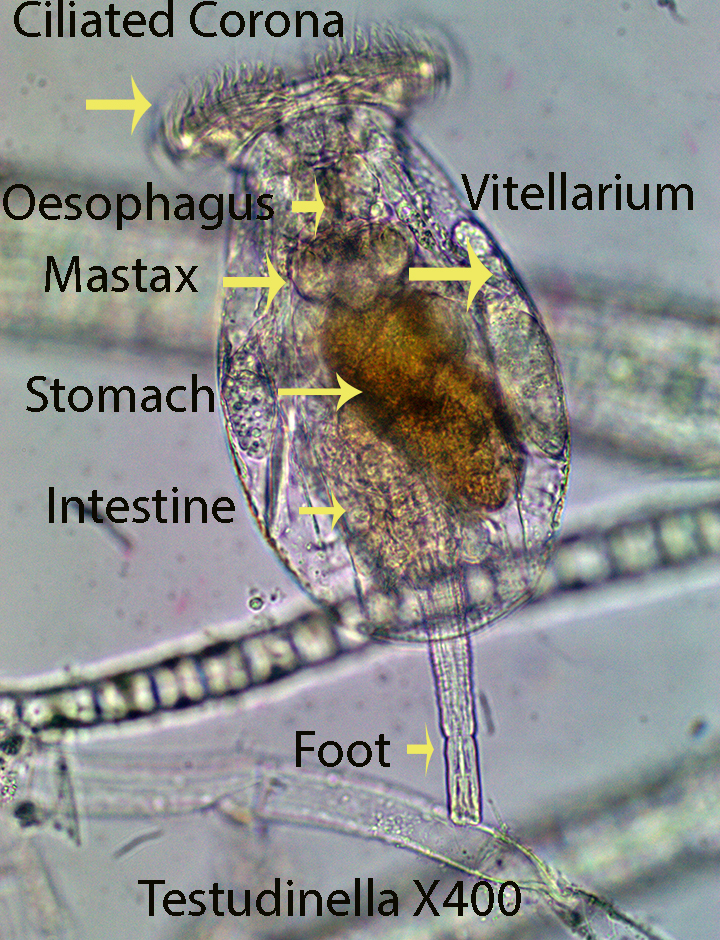
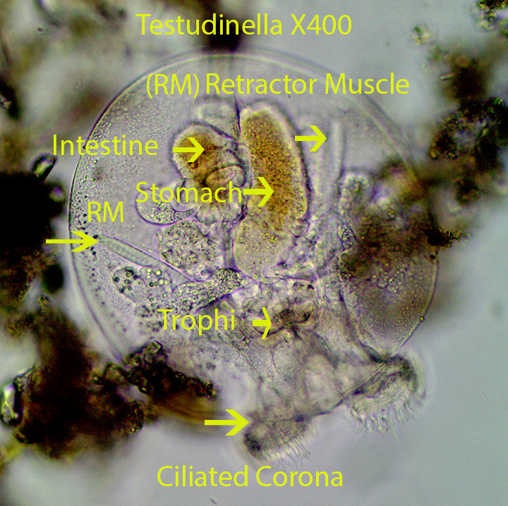
The disc-shaped rotifer , about 325 microns wide, is dorso-ventrally flattened. The ciliated corona can be extended from the protective lorica allowing the animal to move and feed. It also can be pulled into the lorica by retractor muscles during stressful times. The lorica is composed of a dorsal and a ventral plate, fused along the edges. An annulated, ventral , posterior foot, resembling an elephant’s trunk, can be extended from or withdrawn into the lorica. A ciliated cup, at the end of the foot can temporarily attach the rotifer to the substratum during feeding bouts.
https://vimeo.com/162413410 Testudinella X400
https://vimeo.com/118069560 Testudinella X400
29. Trichocera(Trichoceridae)


Trichocera Type A , about 170 microns long, has a ventral mastax with virgate trophi. They possess a simple ciliated corona that is responsible for moving the bean-shaped rotifer from place to place (Refer to the first video). The rotifer often feeds by placing its head directly in front of clumps of debris. The cilia in concert with the mastax drive food (Detritus, bacteria and small protozoans, etc.) into the stomach. Two defining features are: The spine-like toes that are not the same length and a body that is slightly twisted. There are two red eyespots, one of which is visible in the photograph above.
http://vimeo.com/82648785 (Zottoli) (Trichocera X100 Type A Movement)
http://vimeo.com/82648787 (Zottoli) (TrichoceraX400 Type A Anatomy)
http://vimeo.com/85374892 (Zottoli) (Trichocera Type A X400 Anatomy)
http://vimeo.com/85374893 (Zottoli) (Trichocera) Type A X400 Anatomy)
Trichocera Type B, about 300 microns long, has the same basic characteristics as Type A, however it differs in size, shape and other characteristics that are not of concern here. The second video shows this rotifer feeding as discussed for Type A. The third movie shows the rotifer being eaten whole by an unidentified carnivorous rotifer.
http://vimeo.com/85379214 (Zottoli) (Trichocera X400 Type B Anatomy)
http://vimeo.com/85381577 (Zottoli) (Trichocera X400 Type B Feeding)
http://vimeo.com/85379213 (Zottoli) (Trichocera X400 is eaten by an unidentified rotifer)
30. Trichotria (Trichotriidae)


Trichotria, about 450 microns long, is covered completely by a smooth, one piece, rigid lorica. The rotifer which has a mastax with maleate trophi, lacks a circular anterior shield. There are two prominent spines on the foot as shown above.
http://vimeo.com/85390814 (Zottoli) (Trichotria X400 Anatomy)
http://vimeo.com/85390815 (Zottoli) (Trichotria X400 Anatomy)
http://vimeo.com/85390816 (Zottoli) (Trichotria X400 Anatomy)
31. Wulfertia (Proalidae)
Wulfertia is about 160 microns long. The head is not clearly separated from the sac-like body. The frontal corona is reduced in size compared to most rotifers and is covered with short cilia. The rotifer has a small foot with two minute toes. The second movie shows the mastax with its malleate trophi. Feeding was not observed.

Leave a comment