Nematodes
The nematode specimens used to describe male and female anatomy were collected from the fresh water intertidal. This may seem odd, however the basic anatomy of these specimens is essentially the same as those from the bog and the videos are much clearer .
Nematode Movement
The following video shows how nematodes move through soft substrates.
Nematode Anatomy (Male) X400
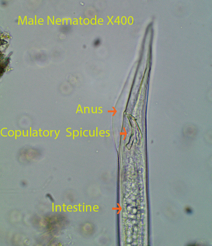
Specimen Description: Initially the video shows the anterior end. The mouth is visible followed by the pharynx. The pharynx joins a muscular, round buccal bulb. It is visible at the bottom of the first screen. Then as the video progresses the connection between the buccal bulb and the intestine comes into focus. The intestinal lining has a greenish tinge to it. The body is surrounded by a cuticle with many circular rings. The posterior end is curved. Note the curved copulatory spicules that lie in a sac that opens through the anus. During copulation the spicules guide amoeboid sperm into the vaginal opening of a female.
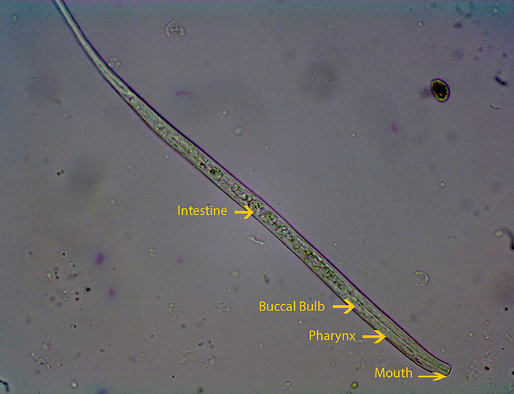
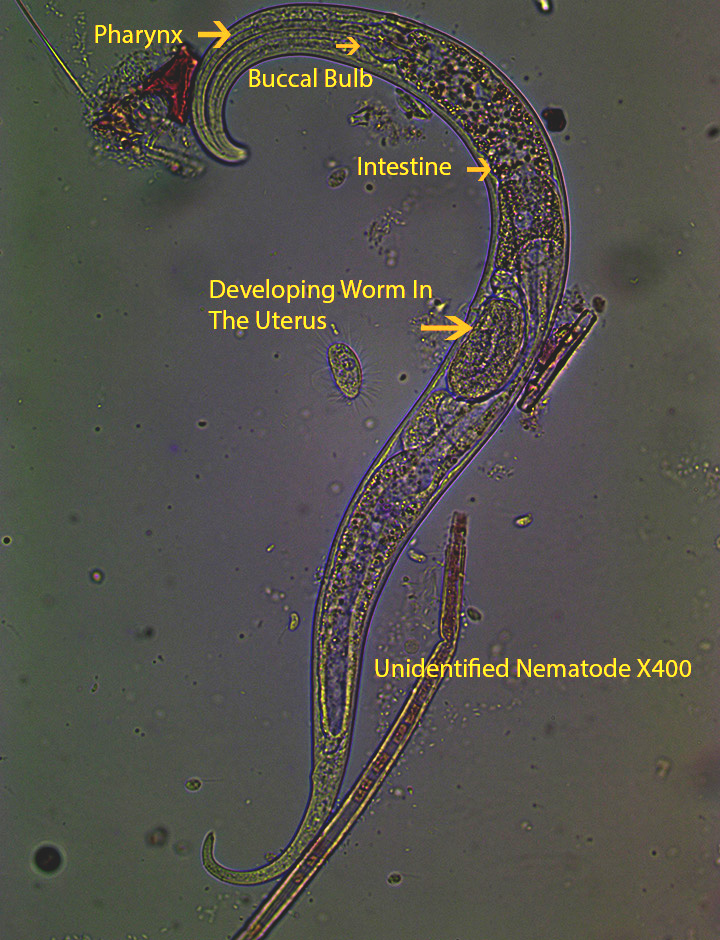
Female Nematode Anatomy – Anterior to Posterior X400
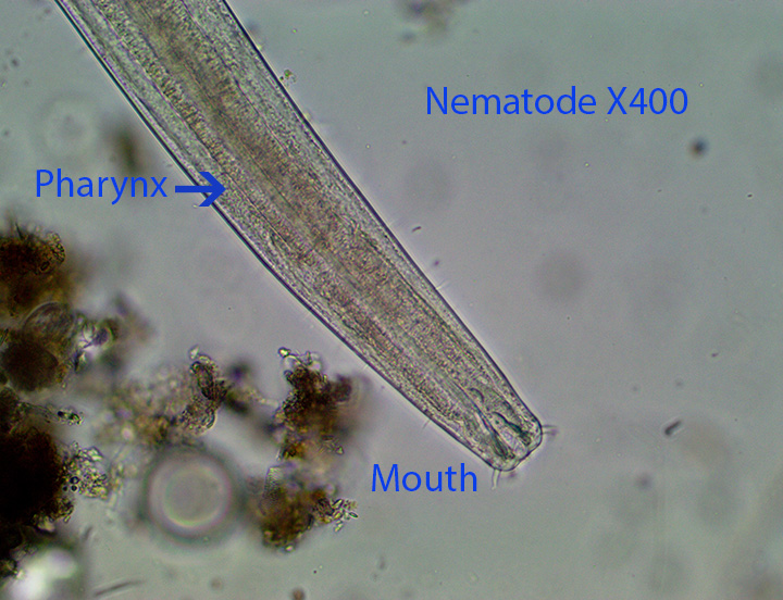
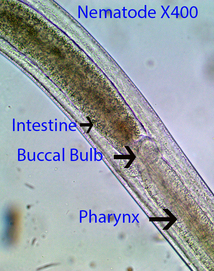
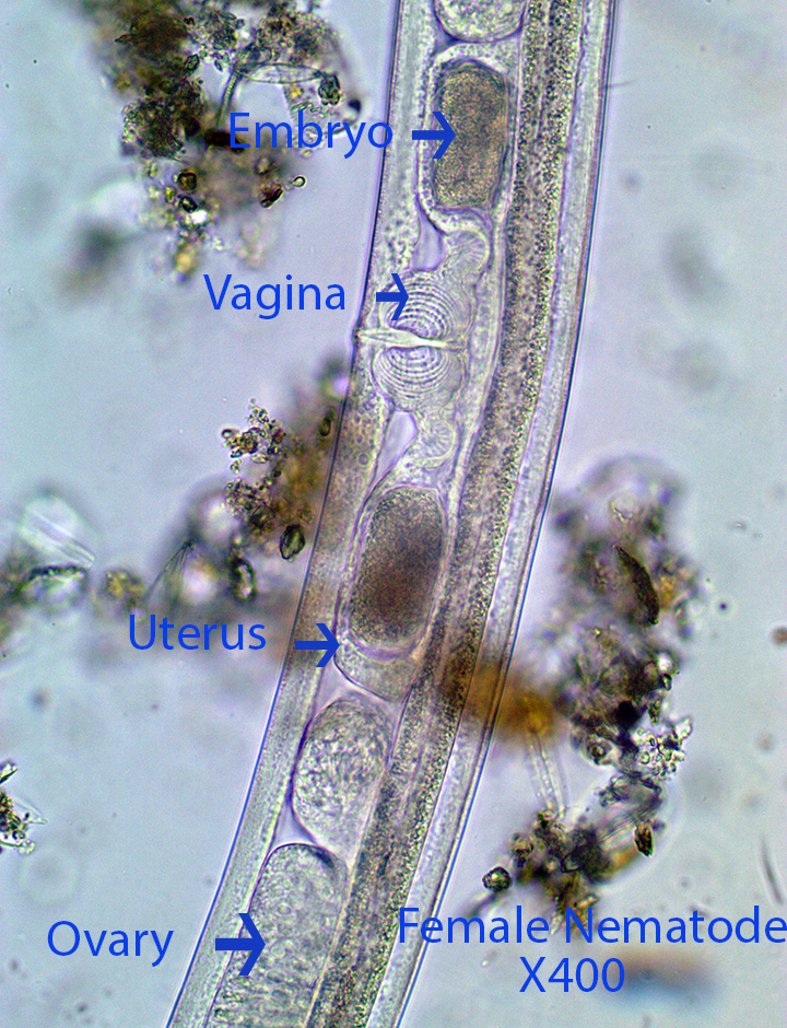
Specimen Description: The female is covered by a ringed cuticle. Thin extensions arise from the cuticle that most likely have a sensory function. The muscular pharynx opens to the outside through the mouth. It is connected to a round buccal bulb that in turn connects to the intestine. The pigmented digestive tract extends backwards almost to the end of the animal where it terminates in a clear triangular cavity that opens to the outside through the anus. The intestine is obscured for the most part by the reproductive system. The reproductive system opens to the outside through a short vaginal canal. The entrance is visible as a slightly raised mound (Vulva) towards the middle of the body; The vaginal canal is located at its center. The vaginal canal splits into two uteri, one projecting anteriorly, the other posteriorly. This is where the fertilized embryos develop. Each uterus connects to an ovary where the eggs are formed. The eggs are almost square with a clear, round, central area that houses the nucleus. A small developing worm is present in each arm of the uterus.
Photographs and Videos of unidentified HVNC Nematodes
http://vimeo.com/86160372 (Zottoli) (Nematode Movement In The Sphagnum Mat X400))
Tardigrades
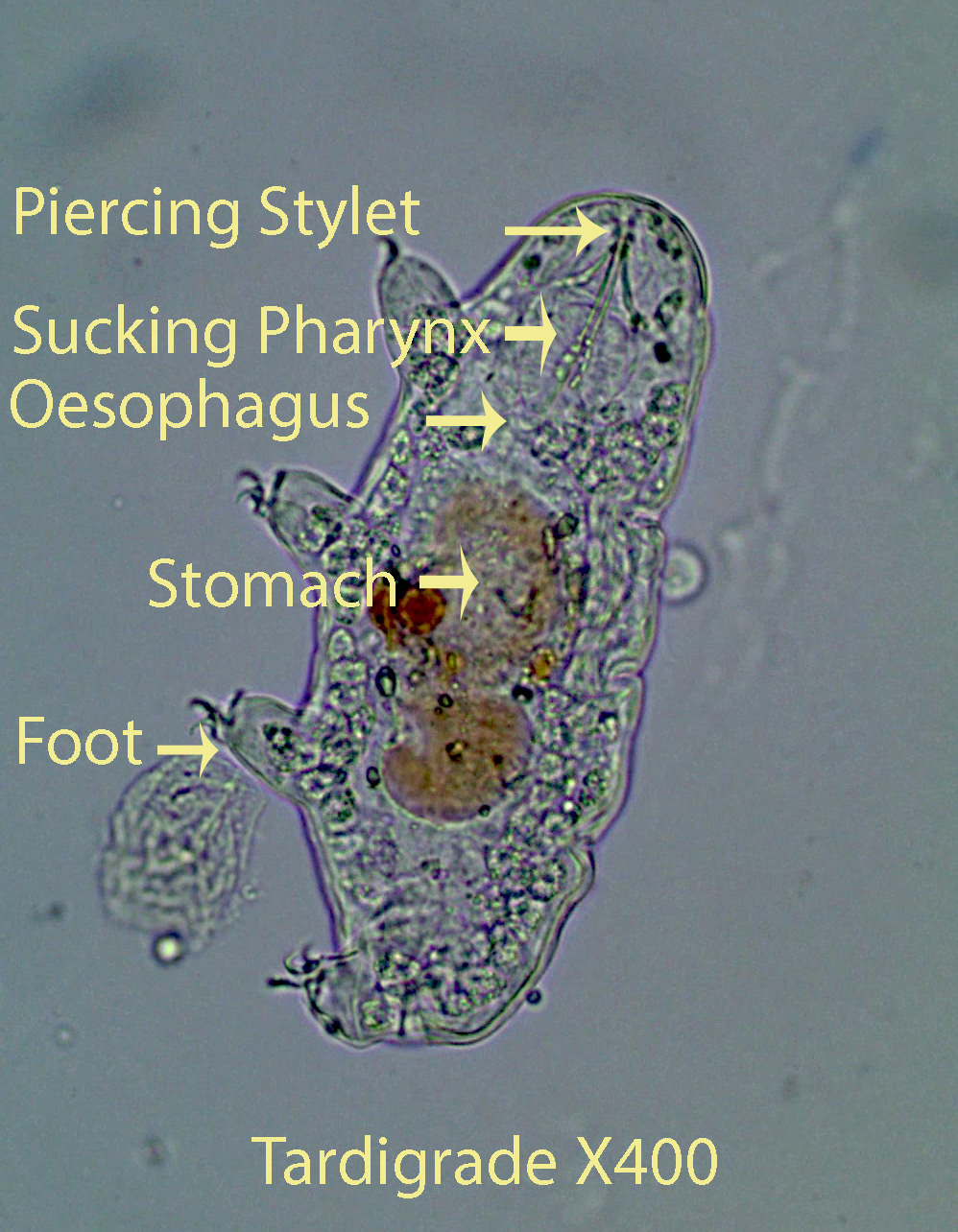
Tardigrades, about 350 microns long, featured in the videos below, most likely belong to the genus Macrobiotus. The animal, cylindrical in cross-section, has four pairs of ventro-lateral, short ,stubby legs. Each leg has two robust double claws and the tips of the claws lie close together. The head lacks cirri and lateral filaments. The anterior mouth leads into a muscular rounded sucking pharynx that connects through a short esophagus to the large colored stomach. In front of the pharynx, note the arrow-shaped piercing stylets that are used to penetrate the cell walls of moss plants and other organisms. Most of these features are visible in the first video. The remaining videos highlight anatomical features as well as movement.
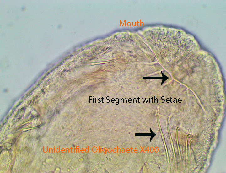
Annelids (Oligochaeta)
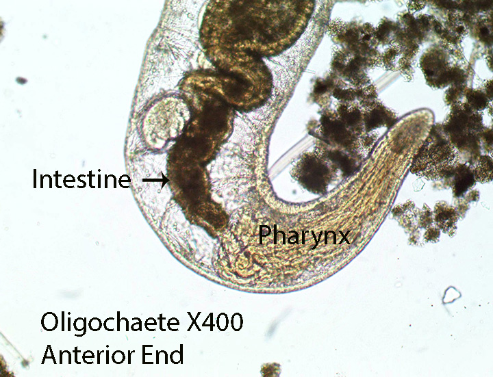
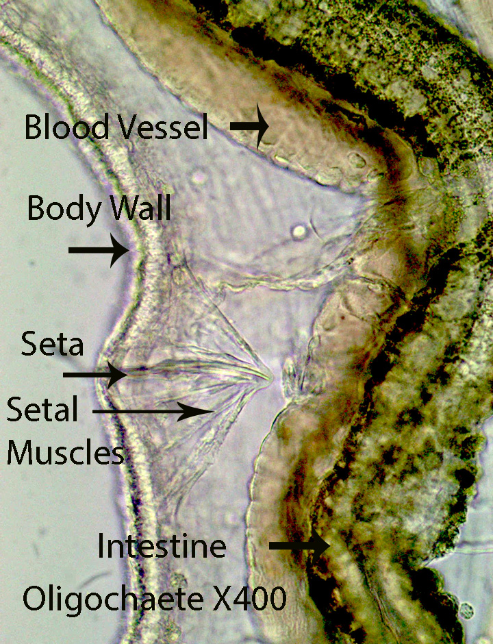
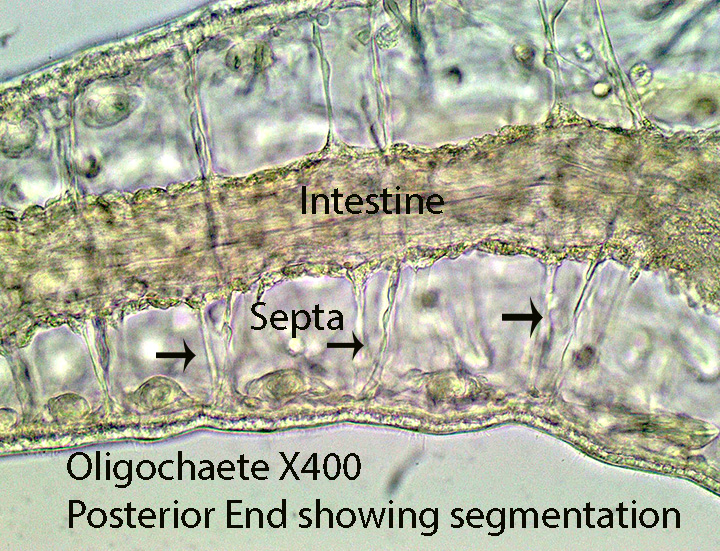
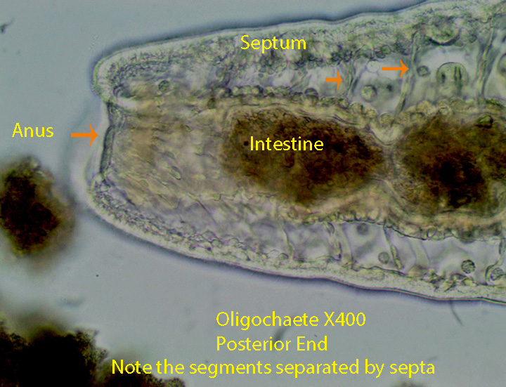
Annelids are multicellular, segmented animals with a mouth and anus. The annelid body is segmented both externally and internally. Externally note the pairs of setae (Pointed Projections) that extend laterally from each segment. They help anchor the body during movement. Internally a sheet of tissue separates each segment at its front and rear boundary. They constrict the digestive tract sometimes making it seem like a “string of beads”. The anterior end is thicker than the posterior and internally bears the orange, muscular pharynx. The dark intestine extends from its junction with the pharynx to the posterior end where food exits through the anus. Peristaltic contractions of circular muscles that surround the gut are noticeable and help move food from anterior to posterior. The red pigment hemoglobin transports oxygen in the blood of most annelids.
Aelosoma(Aelosomatidae)
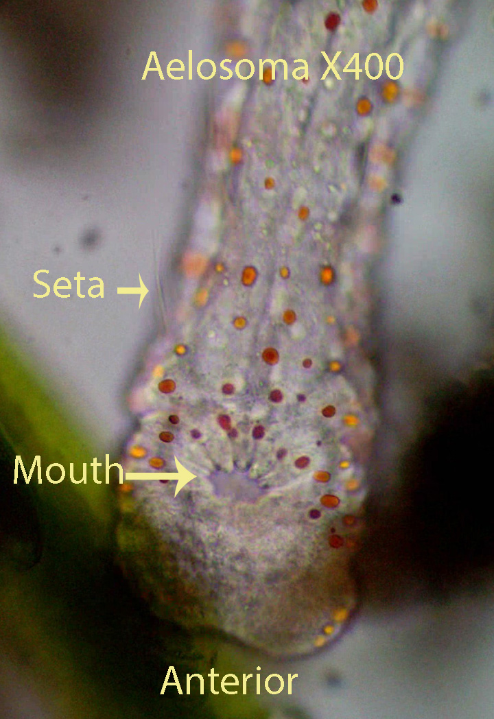
Aelosoma, up to 4 mm long, is characterized by the presence of pigment granules (Red, yellow and green) in the epithelium. The mouth is situated at the back of a rounded head and is connected to a ciliated esophagus. Two dorso-lateral bundles of long capillary setae are found in most segments. Aelosoma reproduces asexually by fission (Forming Zooids).
Pristina (Naididae)
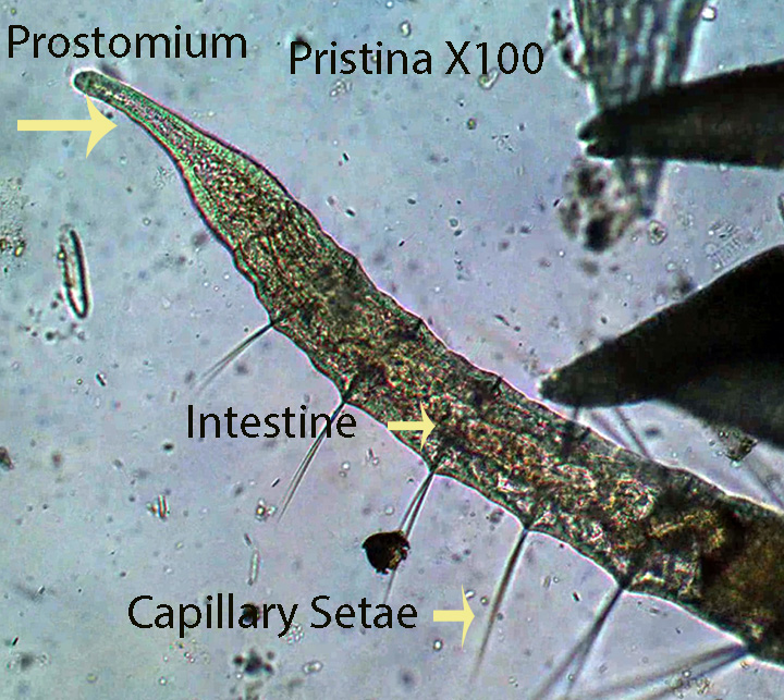
Pristina, about 15 mm long, reproduces asexually by fission (Zooids). The worm lacks colored globules such as those found in Aelosoma. Long, dorsal, capillary setae are present on the second segment as well as on most other segments. The prostomium (most anterior structure) is a relatively long, thin, anterior, finger-like process.


Leave a comment