Arthropods
Arthropods are such a diverse group that I thought it might be useful to discuss some of the characteristics that they have in common before delving into specific groups.Most if not all arthropods are segmented and in primitive forms there may be one pair of appendages per segment.Appendages have evolved in different groups to perform specific functions such as walking, swimming, feeding, and sensing the environment. They generally have a hard exoskeleton that protects them from predators. Once the animal outgrows its confining exoskeleton it must shed (Molt) the old one and form a new one with more room to grow. During this period the organism is in danger of being eaten or physically harmed since the new exoskeleton is initially soft and doesn’t harden for a time. Arthropods generally have an open circulatory system. Blood is pumped from a heart into arteries that pass blood into open sinuses before being returned to the heart. Blood is not enclosed in veins like in our closed circulatory system. Arthropods also have a nervous system consisting of a dorsal brain connected to a double ventral nerve cord.
1. Class Arachnida (The Mites)
Order Prostigmata
Suborder Trombidiformes (Fresh Water Mites)
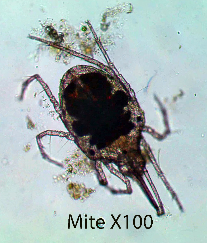
The Suborder Trombidiformes includes mites found in fresh water habitats. There are several species of mites, shown in the videos below, that are common in the Sphagnum mat. The following general description and the videos will at least give you an idea of what a mite looks like. Mites have four pairs of legs, no obvious body divisions, and no abdominal segmentation. The legs have six segments and end in terminal claws.
2. Class Branchiopoda
Order Cladocera
Family Chydoridae
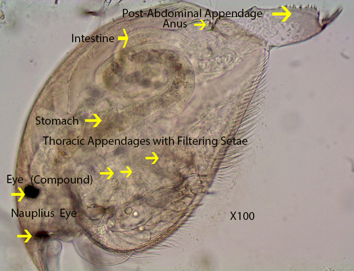
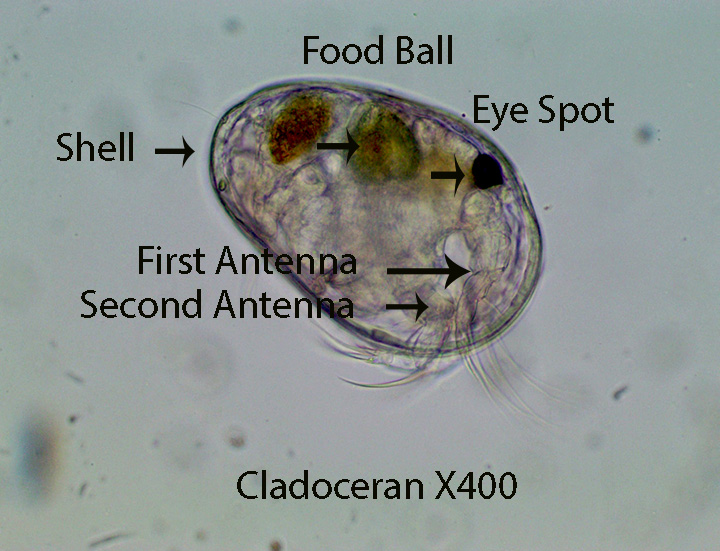
The body, including legs, is covered by a bivalve, opaque, carapace that is attached dorsally. The first pair of antennae (Antennules) are small and partially concealed. The anterior end is elongated into a beak that projects forward in front of the antennules. The second pair of antennae are the longest and are used to move the animal from place to place in the water column. During locomotion, the second pair of antennae are moved upward (Dorsally) and then pulled downward, pushing the animal forward. This sequence is repeated over and over again. Because it takes a short time to bring the appendages upward prior to the downward stroke , movement is jerky. They can also move along the bottom by thrusting their rear post abdominal appendage out of the shell , pushing the animal forward.
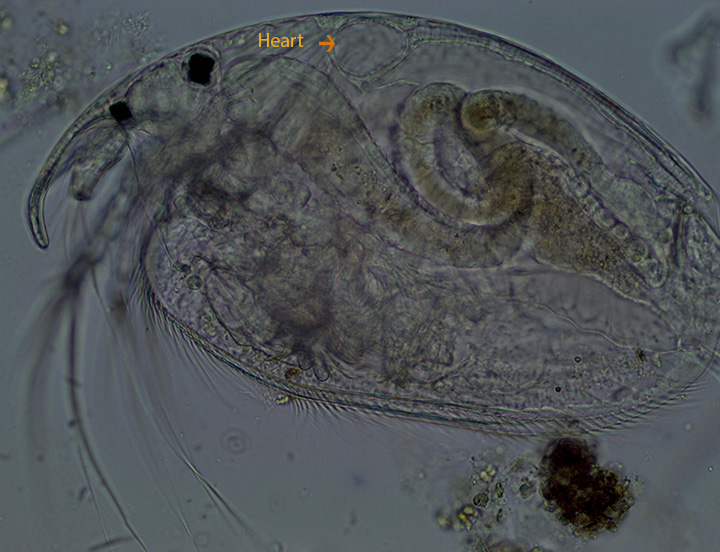
Most branchiopods are filter feeders. The thoracic appendages, enclosed by the carapace, move rhythmically, back and forth, creating a water current that pushes food (mainly detritus, algae and protozoans ) along a mid-ventral groove to the mouth. From here it passes down the esophagus to the stomach and then to the intestine where it exits through the anal opening located dorsally at the base of the post abdominal appendage.. The beating, dorsal heart, located above the stomach, can be viewed in the second video . Most chydorids reproduce parthenogenetically.
http://vimeo.com/86419112 (Zottoli)(Cladoceran X400 Embryos in Brood Chamber)
3. Class Copepoda
The general anatomy of salt marsh pool copepods and those living in the Sphagnum moss bog ecosystem is similar, however, the bog copepods are smaller. I therefore chose to illustrate external anatomy and movement using salt marsh pool specimens described in The Pannes and Pools section of : HTTP://zottoli.wordpress.com/salt marshes/
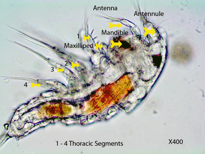
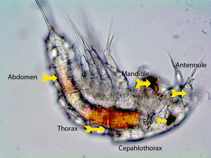
A long (sometimes short) pair of antennules (First Antennae) are visible extending laterally from the head. They are used to propel the animal forward as shown in the second video. The antennules are pulled downward quickly from a dorsal to a ventral position, pushing the animal forward. The antennules are then brought back to a dorsal position and the process is repeated. The thoracic legs also play a part in movement. A single median eye is situated between the two antennae . Thoracic appendages are visible along the wide part of the body. The thorax narrows and joins a thinner abdomen. The cephalothorax, shown above, is the region of the body from the point of the thorax arrow to the right; the thorax is the section of the body from the point of the thorax arrow to the beginning of the abdomen and the abdomen comprises the rest.
Videos
#1 showing the specimen described above:
#2: Movement using antennae (About 4 mm Long)
#3 (Movement): The first part of the video clearly shows a copepod crawling over algal filaments.X400
#4 (Movement): Dorso-Ventral Muscle Contractions used during Movement X400)
#5 (Development) X100. The following movie shows a copepod with a single sac containing developing young, attached to the ventral surface of the abdomen. Some of the young that have escaped the sac can be seen moving slowly using their short antennules.
#6 (Development) X400. The following video shows a nauplius larva that will eventually develop into an adult copepod.
______________________________________________________________
There are two orders of copepods found in the HVNC Bog. Each order is briefly described below.
A. Order Cyclopoida
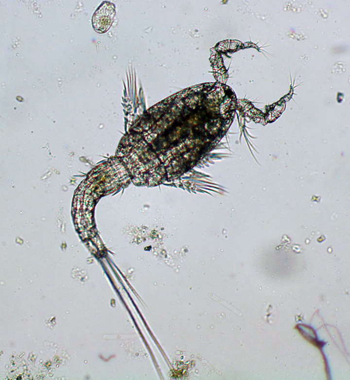

Members of this order have short first antennae, each consisting of 10-16 sections.
B. Order Harpacticoidea
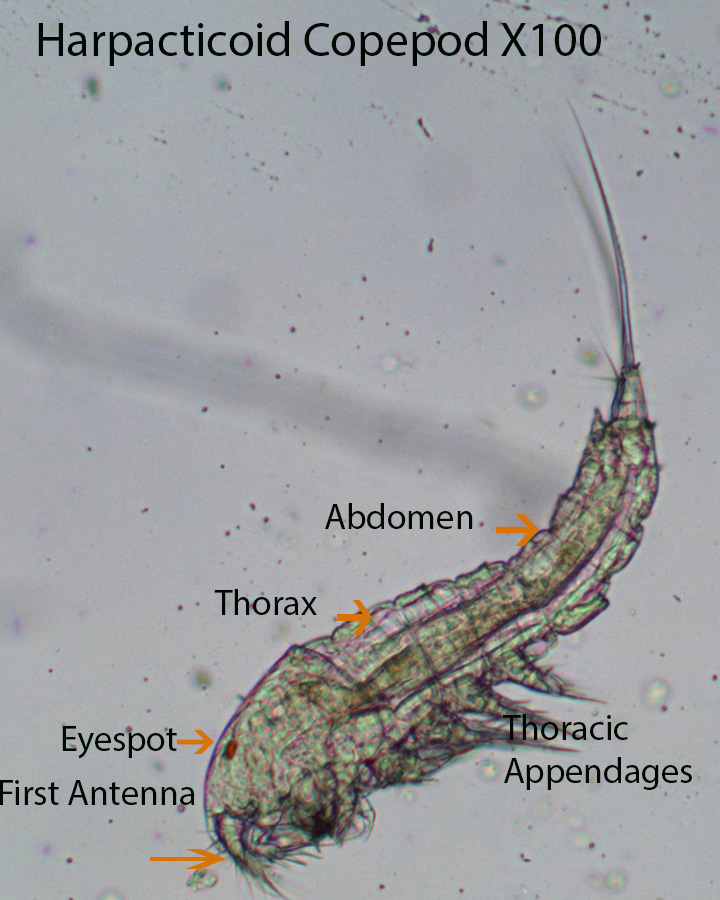
Members of this order have short first antennae (Antennules), each consisting of less than 10 sections.
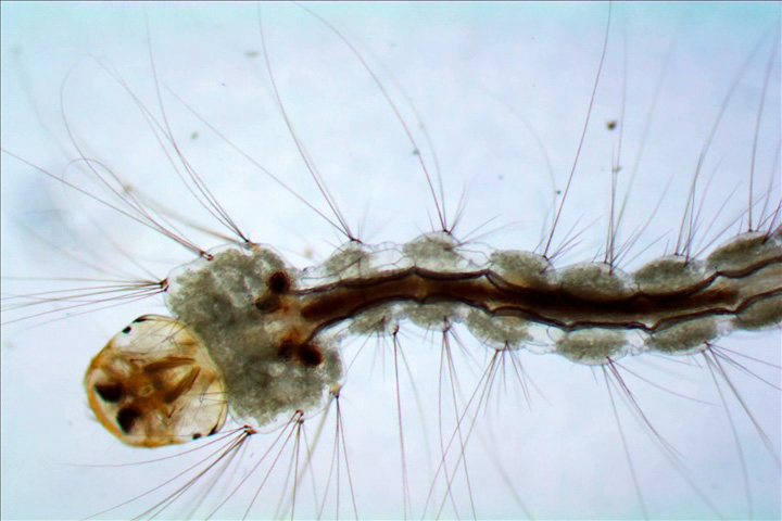
Copepod Nauplius Larva X400 (HVNC Bog)
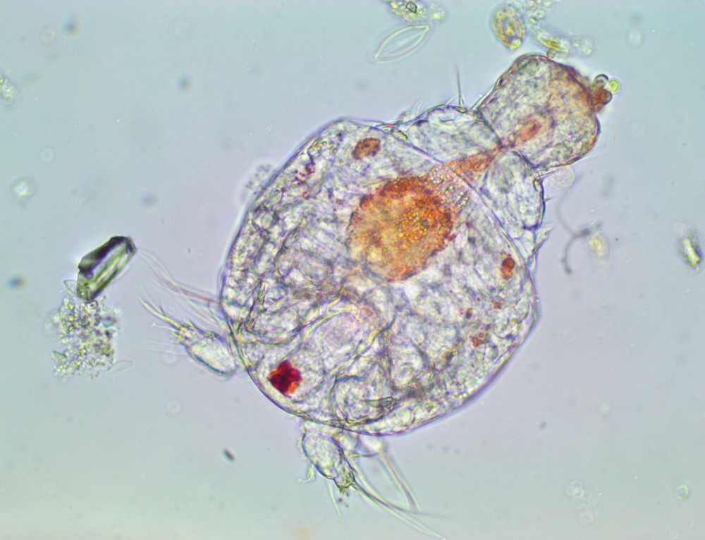
The short, first pair of antennae lie on either side of the red eye- spot. The second pair of antennae are followed by the out of focus mandibles.
4. Class Ostracoda
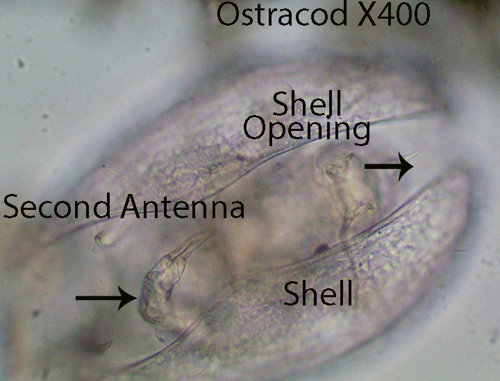
Ostracods are contained within a bivalve carapace (Shell). The body is divided into two regions (Head and thorax). They lack an abdomen. The head bears four pairs of appendages . The first pair (1st Antennae) is generally used for swimming while the second pair (2nd Antennae) ,that trails somewhat posteriorly, helps the animal walk and crawl over the substratum. The third (mandibles) and fourth (Maxillae) pairs are used in the feeding process. The thorax bears two pairs of appendages that are used for walking and crawling as well as for maintaining position. The internal anatomy for the most part is difficult to see and is not discussed here.
5. Class Insecta
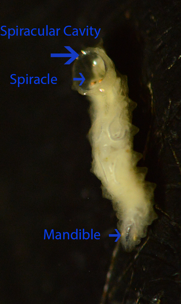
a. Family Ceratopogonidae (Biting Midges)
Alluaudomya spp.

Only one genus of biting midge was found in the sphagnum mat, however I expect there are many more depending on the time of year. Two pairs of black eye spots are visible on the dorsal surface of the sclerotized head capsule (HC). One pair of small antennae project from the anterior portion of the head capsule. Dark mouth parts (Mandibles and labial plate) used for gathering and ingesting food lie within this structure (HC). The ventral labial plate scrapes off detritus, algae and small invertebrates attached to solid surfaces such as algal filaments and the lateral, pointed mandibles push food into the mouth. The mandibles can also directly grab prey. There are 11 segments behind the head. Anterior and posterior pro-legs are absent and long, dark setae project from the posterior end. Striated, longitudinal muscle fibers are visible along the length of the body. The first video documents how the animal moves.
b. Family Chironomidae (HVNC Bog)
1. Pitcher Plant chironomidae Larva (Metriocnemus knabi) X40

Refer to the Invertebrates Living in Pitcher Plants section
c. Family Culicidae
Pitcher Plant Mosquito Larva (Weyomia smithii)
Refer to the Invertebrates Living in Pitcher Plants section.
d. Family Sarcophagidae (Flesh Flies)
Pitcher Plant Sarcophid Blaesoxiph spp.
Refer to the Invertebrates Living in Pitcher Plants section.


Leave a comment