Pitcher Plant
Serracenia purpurea
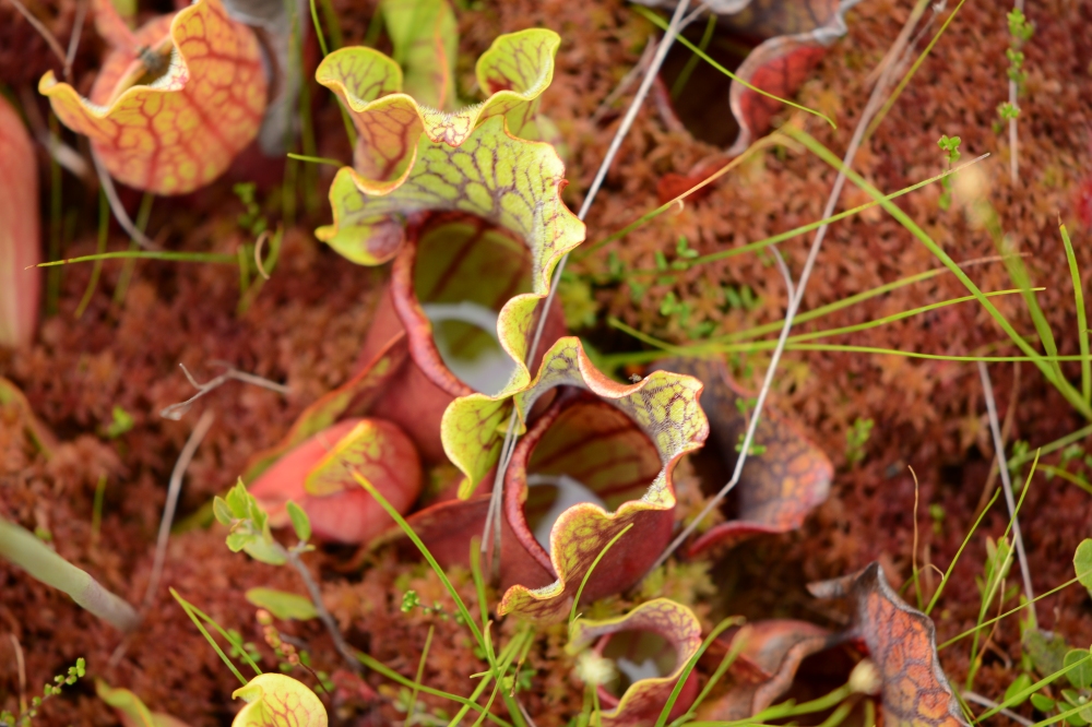
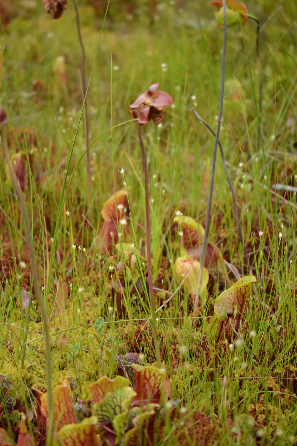
The carnivorous pitcher plant (Serracenia purpurea) consists of a group of vase-shaped, hollow, reddish-green leaves, each about 10 to 30 cm tall, connected to a stem that extends roots downward into the bog. A large flower, supported by a stem arises from the center of the modified leaves as shown above. Several spider species lay their eggs inside this structure. Tea made by steeping leaves in water has been used to treat fever and subdue chills. Native Americans consumed pitcher plant roots to treat liver and lung diseases.
Each “pitcher” has an upper, flared lip that has hairs which curve downward and is generally partially filled with water. Insects (mainly ants) attracted to the “pitcher” crawl inside the modified leaf and are prevented from leaving by the downward pointing hairs and the slippery inner surface of the leaf. Eventually the insects tire and fall into the water where they are digested by bacteria for the most part. The products of digestion, high in nitrogen containing amino acids, are absorbed by the leaf, supplementing photosynthetically produced organic matter. Water contained by the leaf supports a community of interesting organisms that include bacteria, protozoa,, rotifers, chironomid larvae (Metriocnemus knabi) , mosquito larvae (Wyeomia smithii), and sarcophagid larvae (Blaesoxipha) . Videos and descriptions of some of the pitcher plant inhabitants are presented below.
Adaptations of Pitcher Plants:
1. Insectivory: As mentioned above pitcher plants supplement their diet with digested insects.
2. The pitcher plant leaf, described above, is modified to trap and hold insects.
_____________________________________________________________________________________________________________
Invertebrates commonly associated with Serracenia purpurea
1. Phylum Arthropoda, Class Insecta,
a. Chironomid Larvae (Metriocnemus knabi) X40
Family Chironomidae
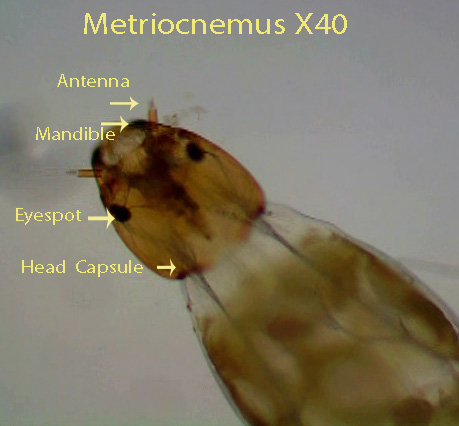
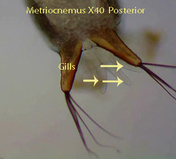
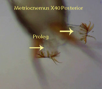
The head capsule is the first visible structure. The two black eye-spots are located laterally on the head followed by two small antennae attached to the dorsal surface of the head capsule. Two, black tipped, lateral mandibles can be seen at the beginning of the video moving back and forth. The labrum (lower lip) lies beneath the mandibles and is used to scrape food (insect remains and plant cells) into the mouth. There are 11 segments behind the head. The first segment bears a single, ventral proleg with numerous spines. It is visible briefly at the start of the video. It is responsible for pulling the animal forward. The last segment gives rise to two prolegs with hooks. Transparent finger-like anal gills are visible underneath the prolegs. A system of visible tubes (tracheae) delivers oxygen throughout the animal. A wide digestive tract runs the length of the larva. The larvae spend most of their time feeding on dead insects lying at the bottom of the pitcher. They may also feed by scraping epidermal cells from pitcher plant leaves.
http://player.vimeo.com/video/24328552
____________________________________________________
b. Mosquito Larva (Weyomia smithii)
Family Culicidae
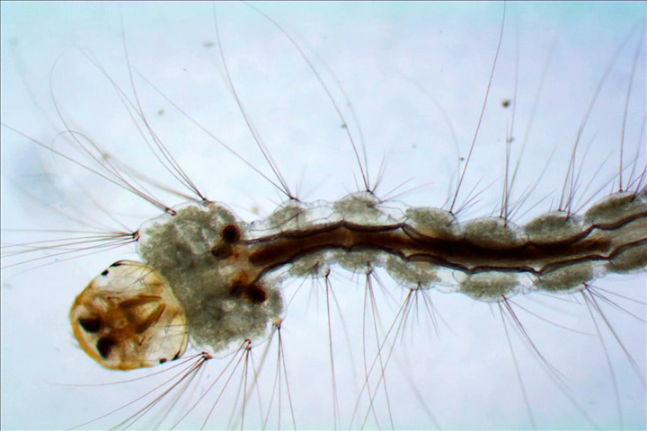
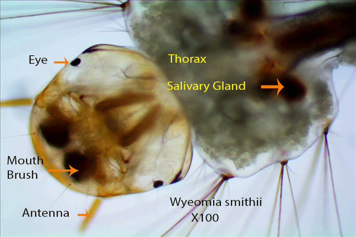
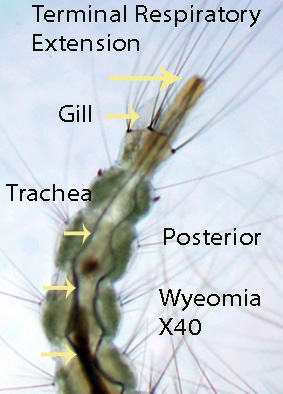
Video A: The posterior end is shown first. Note the terminal respiratory extension on the last abdominal segment. There are two dark tubes within this extension (Tracheal Tubes) that open to the outside. When this extension is pushed above the surface of the water inside the pitcher plant, air containing oxygen moves inward through the tracheal tubes and is distributed throughout the larva. Paddle-shaped gills lie below this structure and are connected to the tracheal system. There are nine abdominal segments. The most anterior is attached to a box-shaped thorax followed by the head capsule. The head capsule has a pair of lateral eye-spots and a pair of dorsal antennae. Internally two dark patches are visible. These are the feeding brushes that can be seen extending outward and then retracted over and over again. The brushes filter food from the water inside the pitcher plant.The swimming larvae are considered top predators feeding for the most part on protozoans and rotifers.
Video B: The same features mentioned above can be viewed here, however the movement of the feeding brushes is easier to see.
VideoA.http://player.vimeo.com/video/24331993
Video B. http://player.vimeo.com/video/24330469
c. Fly Larva Blaesoxipha
Family Sarcophagidae
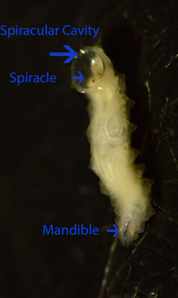
Larvae of this species live exclusively in the fluid filled cup of the pitcher plant Sarracenia purpurea. I have never found more than one specimen in a single cup. They float near the liquid surface feeding on insects (mainly ants) that fall into the pitcher plant before the ants sink to the bottom. In one study, it was determined that Blaesoxipha consumed up to 50% of the insects that fell into pitcher plants. The larval head is incomplete. Note the black mandibles in the photograph and videos. They move parallel to one another as the animal feeds. Blaesoxipha floats, head down, with the deep, posterior, spiracular cavity extending slightly above the surface of the liquid. Two orange, raised ,spiracles lie within this cavity. Air is moved through the spiracles into two large tracheal tubes. The large white tubes, branch into smaller tubes (Visible) that deliver air, containing needed oxygen, to larval tissues. Larvae eventually crawl out of the pitcher plants and pupate on the bog surface.
Bacteria present in the water filled pitcher plant cup form the base of the food chain. They, as mentioned above, are mainly consumed by filter feeding protozoans and rotifers. Mosquito larvae (Top Predator) in turn use their mouth brushes to remove protozoans and rotifers from the water column. Chironomid larvae on the other hand feed on decomposing insects at the bottom of the cup. Finally, the predator Blaesoxipha feeds on dead insects near the water surface as they hang upside down in the water column.
______________________________________________________________________________________________
Phylum Rotifera
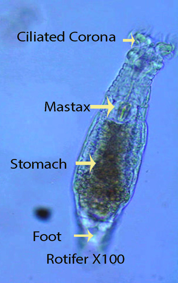
Rotifers are multicellular organisms that can be found in many freshwater and marine habitats. They are temporarily attached to the substratum by a thin foot and two terminal toes. The toes secrete a mucus-like substance that holds them in place. The foot widens into the body (Trunk) proper (Visible) that houses most of the organs. A ciliated bi-lobed disc (Visible) is attached to the body. Disc cilia move the unattached animal through the water column, however, when the animal is attached, the cilia create water movement that brings food (small organisms such as bacteria) into the mouth. Rotifers living inside pitcher plants are filter feeders, consuming bacteria for the most part.Food passes into a muscular bulb called the mastax or pharynx. In most cases the mastax beats (contracts) constantly. The mastax grinds up food and from here it passes into the orange/Brown stomach where digestion takes place. A short intestine follows and empties to the outside through a terminal anus. A clear, round circle (Bladder) can be seen at the end of the trunk near the anal opening. It contracts periodically passing urinary waste to the outside.
A. Video
http://player.vimeo.com/video/24295586
B. Video
http://player.vimeo.com/video/24333020
Note the finger shaped sensory extension from the head
