Ciliated Protozoans
http://en.wikipedia.org/wiki/protozoa
http://en.wikipedia.org/wiki/ciliates
___________________________________________
Table of Contents
A. General
B. Descriptions of Genera accompanied by Videos: Aspidisca, Cinetochilium, Coleps, Colpidium, Dileptus, Euplotes, Frontonia, Halteria, Holophyra, Lacrymaria, Lembadion, Ophrydium, Oxytricha, Paramecium, Paruroleptus, Prorodon, Rhabdostyla, Spirostomum, Spathidium, Stentor, Trachelius, Trichodina, Urotrichia, Vorticella, and Zoothamnion.
__________________________________________________________
A. General:
Protozoans, are generally microscopic, unicellular organisms that are classified by the cellular structures responsible for movement (Pseudopodia, Flagella and Cilia). Protozoans possess membrane bound cellular organelles such as Nuclei, Food Vacuoles, and Lysosomes. Food is captured in many different ways, however in most cases, after capture, it is contained in a spherical bubble (Food Vacuole ) that is formed from the cell membrane and released into the cell interior. Food vacuoles fuse with membrane bound lysosomes that contain digestive enzymes.Enzymes break down food into units that protozoans can use for their own metabolic needs. In freshwater environments, water constantly passes into protozoan cells osmotically. If unchecked, the cell membrane will stretch and rupture. To counter this process most freshwater forms have contractile vacuoles that collect the incoming water and then move it to the outside.
Some of the videos can be used to investigate the anatomy, movement , and feeding mechanisms of select organisms especially in laboratory classes that lack microscopes or funds to buy live animals from biological supply houses. Most students have access to a computer or a cell phone allowing them to reach this site at any time.
B. Descriptions with Videos:
1. Aspidisca (Aspidiscidae)
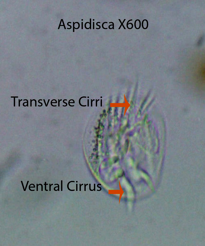
Aspidisca, about 30 microns long, is one of the most abundant ciliates in almost all of the samples collected from April to September. They literally “walk” over the substratum using the fronto-ventral (labeled Ventral) and transverse cirri. The fronto-ventral cirri are more active than transverse cirri and seem to be mainly responsible for movement. In the first video, Aspidisca walks along an Oscillatoria filament filament in search of food. They move over clumps of detritus, then stop for a short time, twitch, and move again. Movement appears to be random. The cell body is inflexible and has four longitudinal dorsal ridges that extend from anterior to posterior. They have an adoral zone of membranelles (AZM) mounted on a stalk (not visible). The AZM is moved over the substratum collecting bacteria, detritus, small protozoans, etc. Bacteria appear to be its main source of food.
http://vimeo.com/81215520 (Zottoli) (Aspidisca)X400
https://vimeo.com/147143476 Aspidisca X600
2. Cinetochilum spp. (Cinetochilidae)
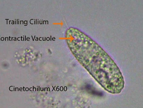
Cinetochilium, about 30 microns long, has a small slit-like, ventral mouth near the anterior end. Cilia are uniformly distributed except for several longer cilia and one trailing cilium that is longer than the rest, both located at the posterior end. One round macro-nucleus is visible in the center of the cell and one contractile vacuole is located near the posterior end. Often numbers of them participate in a “group dance”. Most face upward while rotating on their central, anterior-posterior longitudinal axis. They sometimes rotate from left to right and then reverse direction. At the same time, they wobble from side to side. They are abundant throughout the spring and summer. They appear to be filter feeding on bacteria.
http://vimeo.com/81557729 (Zottoli) (Cinetochilum)
3. Coleps (Colepidae)
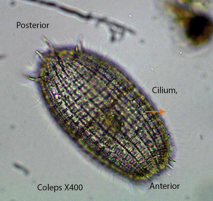
Coleps, about 80 microns long, is covered by external plates secreted by the cell. The barrel-shaped cell body is uniformly ciliated; Cilia protrude through the calcium carbonate plates along visible longitudinal lines. Small spikes surround the anterior and posterior poles. A group of cilia surround the mouth. As the ciliates move forward they rotate from left to right or the opposite way, around their central anterior-posterior longitudinal axis. They also can move forward and backward without rotating. They often push their anterior end into chunks of debris containing bacteria and small algal cells moving slightly from side to side. Oral cilia move dislodged material into the mouth. Micro-tubular rods (not seen) form a circular channel from just below the mouth into the cell interior. They also are attracted to and feed on dead and dying organisms. A single contractile vacuole lies near the posterior end. Coleps is abundant throughout spring and summer. Food vacuoles may contain detritus, bacteria , small algal cells and diatoms.
https://vimeo.com/116174238 Coleps X400 feeding on a Rotifer
https://vimeo.com/147193035 Coleps X400 feeding on bacteria
4. Colpidium(Tetrahymenidae)
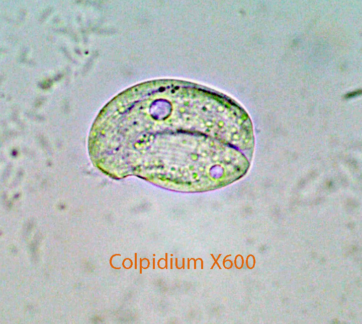
There is an indentation below the anterior part of the cell where the mouth is located. It is most evident in the first video where oral cilia create water currents that bring small particles towards the mouth. Bacteria seem to be the main source of food. The laterally flattened cell body, about 40 microns long, is rounded, pear-shaped, and uniformly ciliated. The front part of the cell is extended slightly to the side. A single contractile vacuole lies in the center of the cell. The cell body of most specimens is filled with food vacuoles.
https://vimeo.com/170318146 Colpidium X1000
5. Cyclidium (Cyclididae)
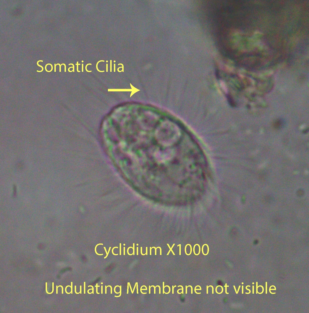
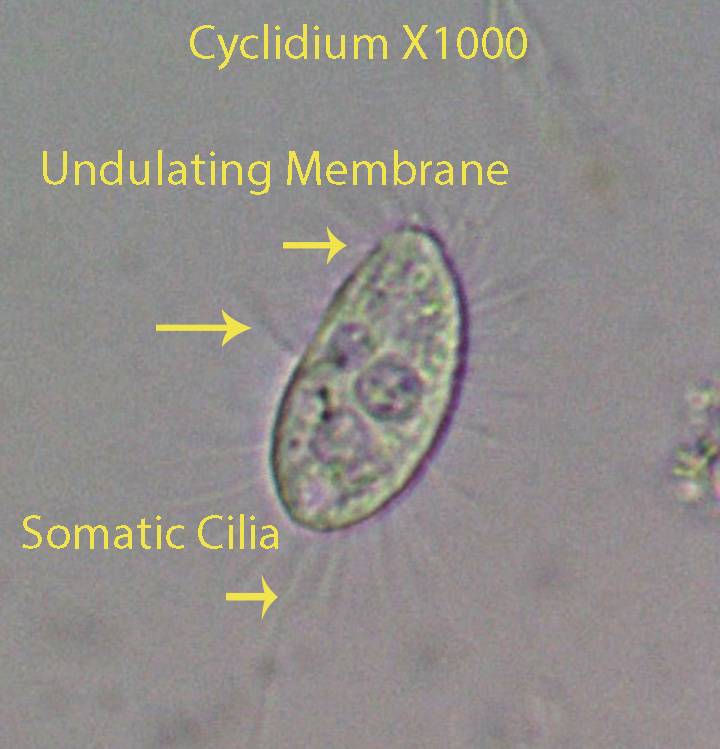
Cyclidium, about 20 microns long, is characterized by the presence of relatively long cilia that extend outward at a 90 degree angle from the cell surface. This is most evident when the ciliate is still, during the feeding process. The cell body is evenly ciliated except for a slight depression towards the anterior end where a transparent sheet-like undulating membrane (Visible). made up of long cilia fused together to form a single transparent sheet. The sheet,extends above the cell surface during feeding bouts. The undulating motion of the membrane directs small particles (mostly bacteria) into the mouth where they incorporated into food vacuoles. If the ciliate is disturbed or stops feeding, they “dart” to a new location usually not far from where they started, and begin feeding again. A single long cilium protrudes from the posterior end. Cyclidium consumes bacteria for the most part. Although the feeding process is visible in this genus it can be seen with greater clarity in the anatomically similar Pleuronema.
![Cyclidium aand nematode-x400-sphagnum-hvnc-july-2013-2x[1]](https://boginvertebrates.wordpress.com/wp-content/uploads/2014/02/cyclidium-aand-nematode-x400-sphagnum-hvnc-july-2013-2x1.jpg)
https://vimeo.com/170474840 Cyclidium X400 Movement
6. Dileptus (Dileptidae)
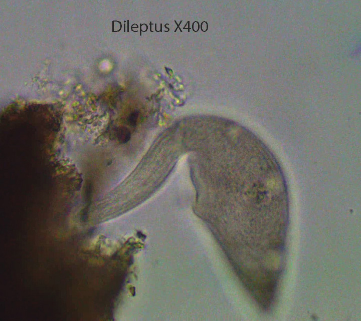
The elongate cell body, about 250 microns long, tapers towards the anterior end, forming a sort of neck. The cell mouth is located ventrally, just behind the neck. The cell body is evenly ciliated. Note how Dileptus rocks back and forth over a clump of detritus. It most likely is feeding on detritus ,bacteria and small protozoans even though it is considered a predator.
http://vimeo.com/81628455 (Zottoli) (Dileptus)
7. Euplotes (Euplotidae)

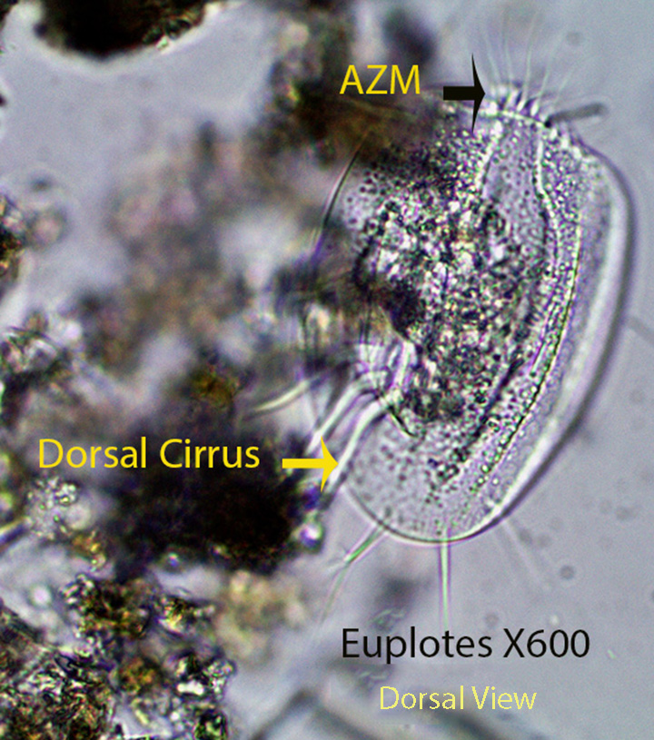
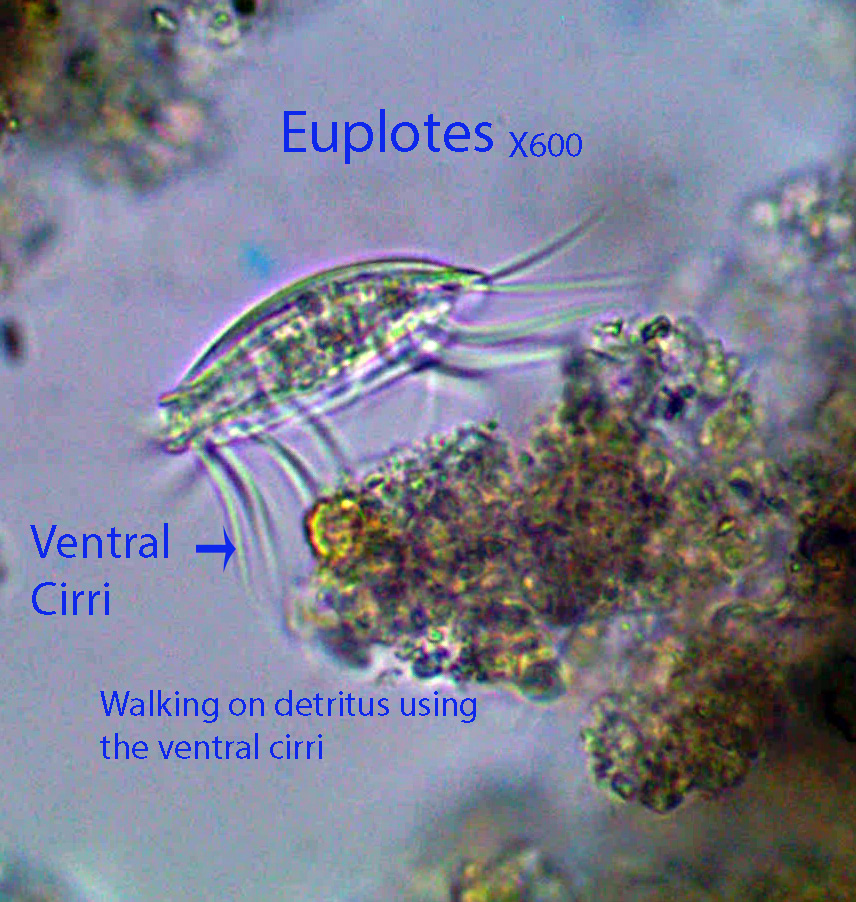
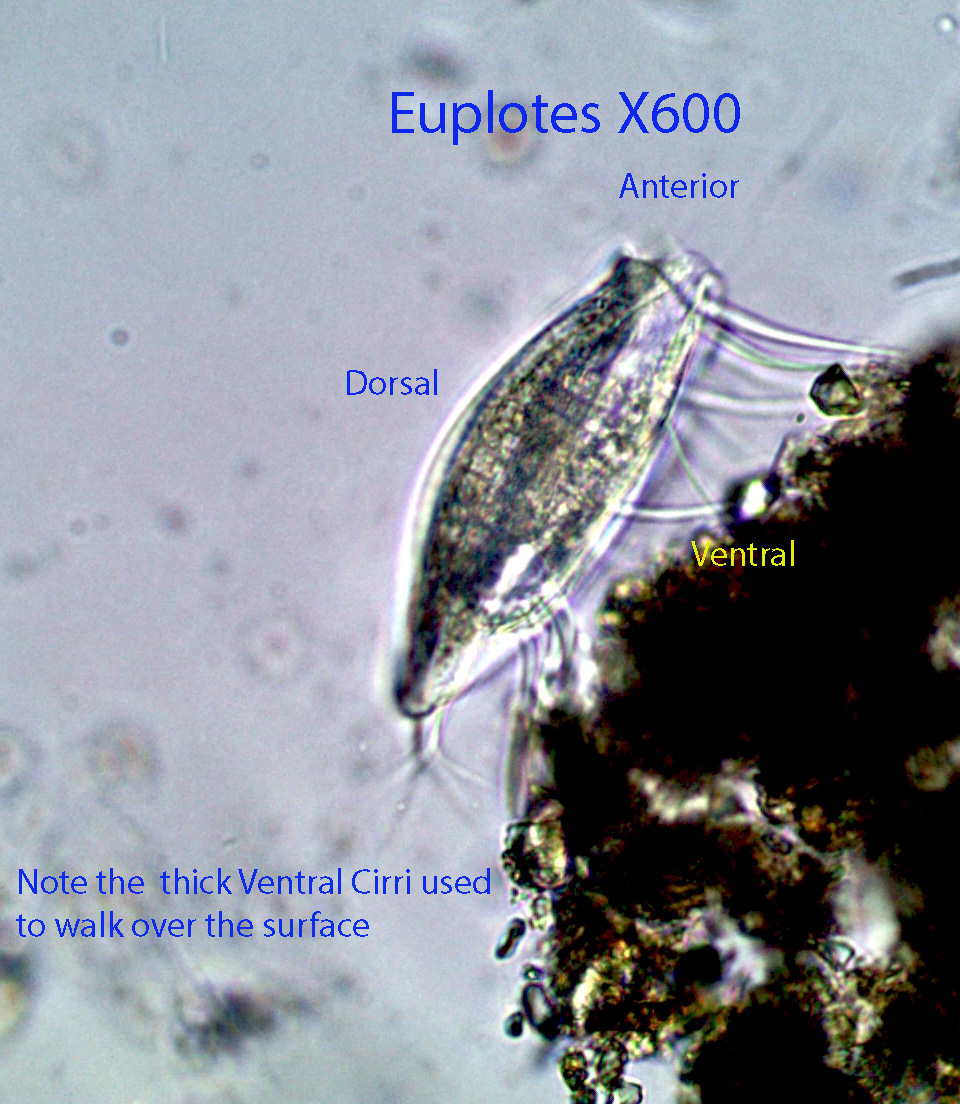
Euplotes, about 50 microns long, has an adoral zone of membranelles (AZM) ,with large cilia that pull water beneath the cell towards the mouth. Suspended particles are removed from the water and pass into the cell interior. They feed on detritus, bacteria, small protozoans, etc. Water circulation is visible in some of the videos. The ventral surface of the cell is flattened and the dorsal surface is rounded (Convex). . The AZM also moves the rigid cell body forward when they travel through the water column. Euplotes has 4 frontal cirri that may have a sensory function, 9 thick ventral cirri, visible in the videos, that are used to walk on the substratum. 4-6 posterior, caudal cirri extend posteriorly from the ventral surface and 5 anal cirri are located at the posterior end; both of these groups seem to stabilize (balance) the animal during movement.
https://vimeo.com/168642111 Euplotes X600
8. Frontonia (Frontoniidae)
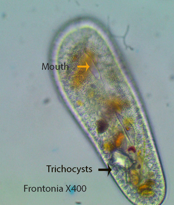
Frontonia is a large (Approximately 400 microns long) oval, dorso-ventrally flattened ciliate with a single layer of vertical trichocysts just underneath the cell membrane. Trichocysts exude filaments that help protect the ciliates from predators. The cell mouth is a small, anterior oval slit shown best in the second photograph. Frontonia feeds for the most part on diatoms and Oscillatoria filaments. A unique green inclusion body, with no known function, is visible in some of the animals. Micro-tubular rods are situated around the mouth. Frontonia is uniformly ciliated and glides smoothly from place to place. They sometimes rotate on their central anterior-posterior longitudinal axis as they move forward. Frontonia is closely related to Paramecium.
http://vimeo.com/81718739 (Zottoli) (Frontonia)
9. Halteria (Halteridae)

Halteria, about 30 microns long, is one of the most common ciliates in the bog. It literally hops from place to place randomly. Locomotion is caused by the quick movement of stiff cilia around the equator of the cell (Visible in the second video). It appears as if they feed when they stop moving. Food is directed toward the mouth by the anterior AZM (Adoral Zone of Membranelles).
http://vimeo.com/27715244 (Zottoli) (Halteria)
https://vimeo.com/52808973 Halteria X400
https://vimeo.com/116179643 Halteria X600
10. Holophyra (Holophyridae)
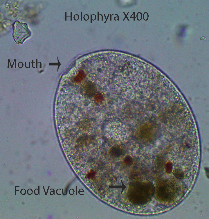
Holophyra, about 300 microns long, is uniformly ciliated. The mouth (Cytostome) is located at the tip of the anterior end. When the animal feeds, the mouth widens as shown in the video, They also feed on smaller prey as indicated by the presence of small food vacuoles containing relatively small particles.The barrel-shaped ciliate Holophyra ,in the first video below, consumes a dinoflagellate. They also feed on diatoms and desmids ,some as long as the animal itself, and small invertebrates. In the second and third videos Holophyra is feeding on a dead copepod.
http://vimeo.com/81720975 (Zottoli) (Holophyra Note the mouth opening at the anterior end)
https://vimeo.com/81720976 Holophyra X400 (Note the diatoms internally)
11. Lacrymaria (Lacrymariidae)

Lacrymaria, about 450 microns long, has a sac-like body with an extensible neck. The fusiform body makes it easier to move into and between clumps of detritus. The mouth (Cytostome) is situated at the tip of the neck. Organelles called extrusomes, located here,release a toxin that kills or quiets prey making them easier to ingest. The ciliate spends most of its time embedded in debris moving its neck back and forth in search of food. The neck can be extended to about twice the length of the body. Somatic cilia are arranged in spiral rows that are most evident on the posterior part of the cell body. They feed for the most part on small protozoans.
http://vimeo.com/74466814 (Zottoli) (Lacrymaria)
http://vimeo.com/81723650 (Zottoli) (Lacrymaria)
12. Lembadion (Lembadionidae)
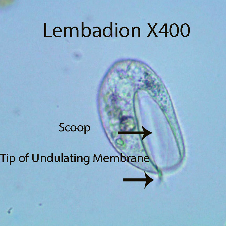
Lembadion, about 95 microns long, is scoop-shaped. There is an undulating membrane within the scoop that can be seen moving back and forth as the animal moves forward. The cell body rotates around the anterior-posterior axis as it moves forward. The tip of the undulating membrane extends slightly beyond the front of the scoop. The scoop might help the ciliate hold prey in place as they are forced into a food vacuole. Small flagellates have been observed inside food vacuoles.
http://vimeo.com/81726343 (Zottoli) (Lembadion)
13. Ophrydium ( Ophyridaceae)
http://en.wikipedia.com/wiki/ciliate (Ophrydium)

The ciliated protozoan Ophrydium spp.,about 300 microns long, has a crown of cilia around its anterior end. The cilia create a current of water that directs particulate matter in the water column towards the “mouth” located in the center of the crown (Filter Feeding). Food then passes down a short gullet into a football-shaped food vacuole (Visible). The small dots inside are probably bacteria; however larger green flagellated protozoans are common prey. The single food vacuole breaks free and drops into the cytoplasm (Visible) and fuses with membrane bound lysosomes (Not Visible) that contain digestive enzymes. This process is clearly visible in the movie. The enzymes break down food into units that the protozoans can use for their own metabolic needs .
http://vimeo.com/81539034 (Zottoli) (Ophrydium spp. Feeding)
14. Oxytricha (Oxytrichidae)

The dorso-ventrally flattened Oxytricha has an anterior adoral zone of membranelles (AZM) on the right side and an undulating membrane made up of long cilia fused together to form a single transparent sheet,on the left. The undulating membrane creates water movement that draws small and medium size particles (Detritus, bacteria, diatoms and small protozoans, etc.) towards the mouth (Cytostome) where they may be consumed. The AZM extends from the mouth, along the left side and across the anterior end. The ciliate, about 150 microns long, has two marginal (along the edge) rows of cirri that are continuous around the posterior end. Other cirri however are not arranged in rows. Ventral cirri allow the animal to “walk” on the substratum. As the ciliates move forward they may suddenly jerk backwards and then move forward again. This process may be repeated again and again. The dorsal surface is somewhat rounded.
http://vimeo.com/81728650 (Zottoli) (Oxytricha)
15. Paramecium (Parameciidae)
http://en.wikipedia.com/wiki/Paramecium

Paramecium is about 230 microns long. Trichocysts are visible as short vertical lines just underneath the cell membrane. When the ciliate is disturbed, a long thin sticky thread is released from some or most of the trichocysts. Mass release of these filaments make it difficult for protozoan predators to ingest them. This defensive function has recently been confirmed experimentally. A small number of threads released from the same area may also allow the ciliate to temporarily attach to the substratum.
A pre-oral groove, visible in some of the videos, leads from the anterior end to the mouth. The mouth is a circular opening that leads to a short tube (Gullet or Cytopharynx) that empties into a food vacuole. The food vacuole eventually fills with prey,detaches, and moves into the cytoplasm. Food vacuole formation can be observed in the following video: http://vimeo.com/82801688 (Zottoli). A new food vacuole forms as soon as the old one leaves. Cell organelles called lysosomes, filled with digestive enzymes, fuse with food vacuoles, dumping their load into the vacuole interior, initiating the digestive process. After digestion is complete the food vacuole merges with the anal pore and its contents are released outside the cell as shown in the following video: http://vimeo.com/82801689 (Zottoli). The cytoplasm moves inside the cell (Cyclosis) as shown in several of the videos. Paramecium living in freshwater take in water osmotically since the concentration of dissolved substances is greater inside the cell than outside. Organelles called contractile vacuoles, one at each end of the cell, receive excess water from radiating canals and squirt it outside the cell when they contract. This process can be observed in the last video.
http://vimeo.com/82801688 (Zottoli) (Paramecium)
http://vimeo.com/82801689 (Zottoli) (Paramecium)
http://vimeo.com/81728652 (Zottoli) (Paramecium)
http://vimeo.com/84401028 (Zottoli) (Paramecium X400. Note the circulating cytoplasm and the posterior contractile vacuole with its star-shaped water collecting ducts)
16. Paruroleptus (Oxytrichidae)

Paruroleptus, about 350 microns long, has two straight rows of marginal (along the edge) cirri as well as two rows of mid-ventral cirri arranged in a zig-zag pattern. The flexible ciliate has an adoral zone of membranelles (AZM) that create water flow towards the mouth. After entering the mouth food particles are directed down to the base of the cytopharynx where a food vacuole is formed. They feed on bacteria, diatoms and small protozoans. The cell body narrows at the posterior end forming a sort of tail.
http://vimeo.com/81715963 (Zottoli) (Paruroleptus X400)
http://vimeo.com/81716288 (Zottoli) (Paruroleptus X400)
17. Prorodon (Prorodonidae)

Prorodon, a ciliated protozoan, is about 100 microns long. It has an apical mouth (Visible) with internal micro-tubular rods (Visible) that help the ciliate manipulate food. In the first movie Prorodon attacks a dead member of its own species. The mouth gradually widens as it is pressed against the cell membrane of its prey. Eventually the animal is drawn inside.
http://vimeo.com/114166244 Prorodon feeding on another Prorodon X400
http://vimeo.com/82142966 Zottoli (Prorodon Feeding on a Dead Rotifer)
18. Rhabdostyla (Epistylidae)

Rhabdostyla , a solitary ciliate, about 40 microns long, is attached to the substratum by a short non-contractile stalk. Cilia are reduced to a band around the anterior end. They often attach to invertebrates such as ostracods and cladocerans. The ciliates filter the surrounding water, feeding on detritus, bacteria, and small protozoans, etc.
http://vimeo.com/83936023 (Zottoli) (Rhabdostyla)
http://vimeo.com/52809371 (Zottoli) (Rhabdostyla)
19. Spirostomum (Spirostomidae)
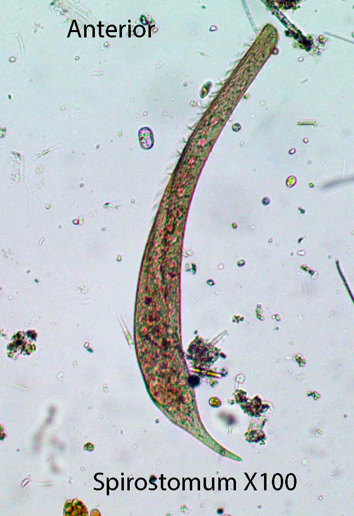
Spirostomum is a long, thin ciliate (Up to 1 mm in length) with a distinct, large, round, clear contractile vacuole at the posterior end. The anterior end of the cylindrical ciliate is narrower than the posterior. The uniformly ciliated cell body is extremely flexible as it navigates over and under obstacles in its path. The animal can move smoothly in a straight line as well as twist and turn as it navigates through debris. Often it will move forward a short distance and then backward and forward again. An adoral zone of membranelles (AZM) leads from the anterior end to the cytostome (Cell Mouth) located where the narrow anterior abruptly widens (About 1/3 of the way towards the posterior end). The body is uniformly ciliated and rows of cilia are visible. The macro-nucleus, visible in some of the specimens, looks like a “string of sausages.” It appears to feed on detritus, bacteria and small protozoans.
http://vimeo.com/83937838 (Zottoli) (Spirostomum)X400 Anatomy
http://vimeo.com/81733245 (Zottoli) (Spirostomum) X100. Movement
http://vimeo.com/81733230 (Zottoli) (Spirostomum) X400. Anatomy
http://vimeo.com/81733228 (Zottoli) (Spirostomum) X100 X400 Movement
20. Spathidium (Spathidiidae)
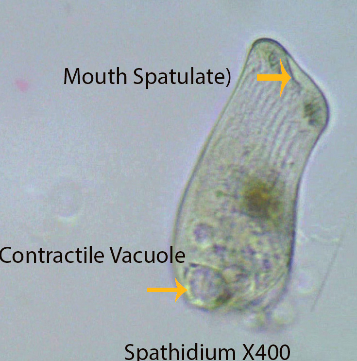
Spathidium is a sac-shaped, flattened ciliate about 150 microns long. The cell mouth is relatively wide (Spatulate) and bears a ring of cilia around its edge. The oral cilia are responsible for moving the body as well as directing food into the mouth. They also rotate around their anterior-posterior axis as they move forward. Extrusomes located around the oral opening help to kill and trap prey making it easier for the ciliate to ingest them. Spathidium is a predator that feeds on medium-sized protozoans and rotifers.
http://vimeo.com/81557728 (Zottoli) (Spathidium)
21. Stentor (Stentoridae)

Stentor, about 1 mm long, is characterized by its trumpet-shaped body. The anterior end is widest and bears an adoral zone of membranelles which create a water current that directs food towards the mouth. The posterior, thinner end, that is attached to the substratum, serves to hold the animal in place. The ciliate can detach the posterior end and move to a new location. Somatic cilia cover the entire cell body.
https://vimeo.com/115068795 Stentor X600
22. Trachelius spp. (Tracheliidae)
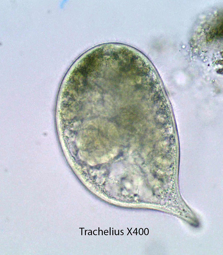

Trachelius, about 150 microns long, is distinguished by a short tapering neck and a round cell body. The round cell mouth (Cytostome) leads into a short tube (Cytopharynx) that ends internally. Food vacuoles are formed at the end of the cytopharynx. The Y-shaped structure (mouth and cytopharynx) are visible in the video below.
https://vimeo.com/81210133 (Zottoli) (Trachelius X400,) of a specimen collected in a local bog.
The animal rotates around its central anterior-posterior longitudinal axis. Feeding was not observed, however the animal is a known predator.
http://vimeo.com/81213412 (Zottoli) (Trachelius X100, Movement)
http://vimeo.com/81210133 (Zottoli) (Trachelius X400, Internal Anatomy)
23. Trichodina spp. (Trichodinidae)

Trichodina lives as an ecto-commensal on the surface of Chlorohydra (Phylum Cnidaria). The first part of the movie shows the body and tentacles of Chlorohydra. Note the internal distribution of the endo-symbiotic green algae. Oral cilia on the anterior (Upper) end are responsible for creating water movement towards the mouth allowing the animal to consume (Filter Feed) organisms such as bacteria and small protozoans as well as detritus. Posterior cirri grasp and pull the ciliate down on the epidermis of Chlorohydra possibly creating suction that might help the ciliate maintain its position while feeding. I have seen Trichodina moving quickly over the tentacles and body of Chlorohydra.
http://vimeo.com/53785031 (Zottoli) (Trichodina)
24. Urotricha spp. (Urotrichidae)
Urotricha, about 20 microns long, has a mouth (Cytostome) with a small circle of slowly beating cilia around the mouth opening. Slowly beating Cilia are also present on the cell body. A single, long cilium extends from the posterior end. The ciliate can expand the apical mouth opening to accommodate small protozoans at least as large as itself.
http://vimeo.com/84000602 (Zottoli) (Urotricha X400) Note the slow beating cilia on the body and around the mouth as well as the empty, round food vacuoles). The single long cilium is visible several times in the video, however it is difficult to see.
25. Vorticella spp. (Vorticellidae)

Vorticella is similar anatomically to Zoothamnion described below except that it is not colonial. One cell, about 50 microns long, is attached to one contractile stalk.The cilia create a current of water that directs particulate matter in the water column towards the “mouth” located in the center of the crown. Food then passes down a short gullet into a football-shaped food vacuole (Visible). The small dots inside the vacuoles are probably bacteria.
http://vimeo.com/84062489 (Zottoli) (Vorticella X400. The following anatomical features and processes can be clearly observed: Food Vacuoles both large and small; Movement of cytoplasm within the cell; Contractile Stalk; and Cilia). Note the food vacuole forming on the right upper side and watch how it separates and moves downward.
26. Zoothamnion spp. (Zoothamniidae)

Zoothamnion is a colonial ciliate with two or more cells (Each about 50 microns long) that are attached to a single contractile stalk. The stalk is attached to the substratum. It is not unusual to see Zoothamnion colonies attached to arthropods such as cladocerans and ostracods.When disturbed ,the entire colony contracts at the same time. A contractile thread in the center of the stalk reaches every the cell in the colony. When the thread contracts the stalk pulls the colony downward. Cilia form a band around the anterior end of each cell. Water is drawn by these cilia from beneath the cell towards the top of the cell body and back down in a circular path. Food particles tend to drop to the center and may be directed into the mouth (Cytostome),down the cytopharynx and into a food vacuole. Food may include detritus, bacteria and small protozoans.

Such a greaaaaaaaat ressource ! Much respect ! R
Tremendous site. I find it accidently and immediately identified (as a lembadion) something I’d known just as ‘scoop creature.’