Cnidaria (Coelenterates)
Chlorohydra (Hydridae)

Chlorohydra, about 5 mm long, is a radially symmetric animal with a sac-like body. A circle of 5 tentacles originates from the anterior end. The mouth is located in the center of a dome-shaped mound of tissue (Hypostome) located above the base of the tentacles. The mouth leads into a blind ending sac (Gastrovascular Cavity or Gut) where digestion takes place. The digestive tract has only one opening that serves as both mouth and anus. Tiny stinging cells (Nematocysts), concentrated in tentacles, immobilize prey such as small cladocerans. Prey are then directed by tentacles into the mouth and then passed into the gut where digestion takes place. Later hydra ejects undigested remains through the mouth opening. The body wall is made up of two layers (Ectoderm and Endoderm) separated from each other by the non-cellular mesoglea. Green, single cell, algae (Zoochlorellae) (Visible) live inside endodermal cells (Nutritive-Muscular Cells) lining the gastrovascular cavity. A symbiotic relationship exists between the Zoochlorellae and Chlorohydra. Algae provide photosynthetically derived carbohydrates and oxygen to the host, while the host provides shelter and a source of carbon dioxide needed in the photosynthetic process. The ectoparasitic ciliate Trichodina spp., described in the protozoan section, is visible on the external surface of hydra.
Platyhelminthes (Flatworms)
All of the genera below belong to the class Turbellaria. They are elongated animals that are generally flattened dorso-ventrally and have cilia on most body surfaces. Flatworms have a digestive tract with one opening that serves as both mouth and anus. The mouth is usually positioned ventrally, however it can be terminal or sub-terminal. The digestive tract begins with a muscular pharynx that connects to a digestive tube. Platyhelminths are hermaphrodites capable of reproducing sexually. Three of the genera (Catenula, Rhynchoscolex and Stenostomum) discovered in the HVNC bog are able to asexually form chains of individuals (Zooids) that are genetically identical to the parent. The zooids eventually separate from the parent and go their own way. Eye-spots may or may not be present. Cilia, on the flattened ventral surface propel the animal smoothly over the substratum, however many species with well-developed body cilia can swim freely throughout the water column.
Catenula (Catenulidae)
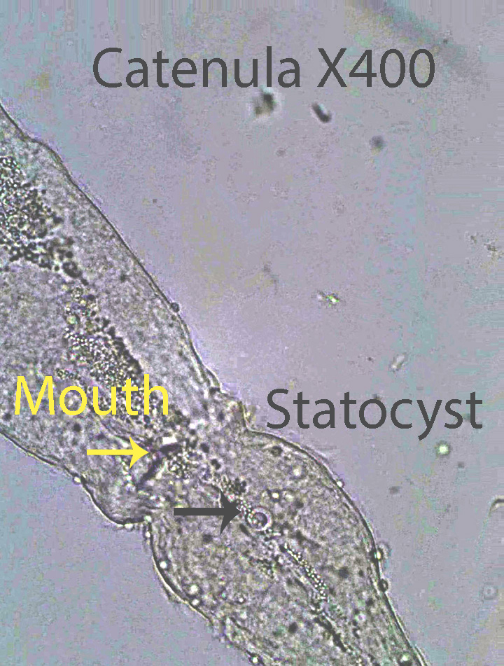
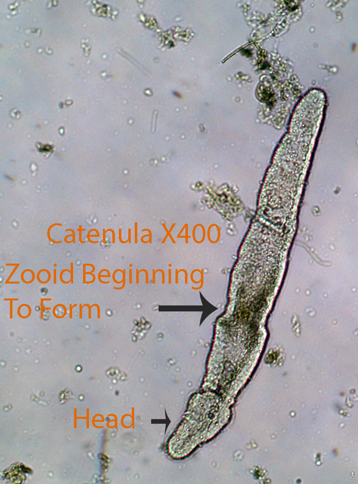
Catenula, about 1.75 mm long, has a rounded head region separated from the rest of the body by a circular groove. The relatively small flatworm has a characteristic, round, statocyst on the head region. A ventral, round mouth opening is located behind the head. It leads into a sac-like digestive tract. Catenula is often divided into a series of zooids. During active feeding bouts they may twist and turn using mesoderm derived, circular and longitudinal muscles.
Microdalyellia (Dalyelliidae)

Microdalyellia, about 1 mm long, has a cylindrical body with a rounded anterior and pointed posterior. The mouth is located apically and leads into a large, muscular, bulb-shaped pharynx connected to a sac-shaped digestive tract. There are two anterior eye-spots. During active feeding bouts they may twist and turn using mesoderm derived, circular and longitudinal muscles.
Rhynchosolex(Stenostomidae)
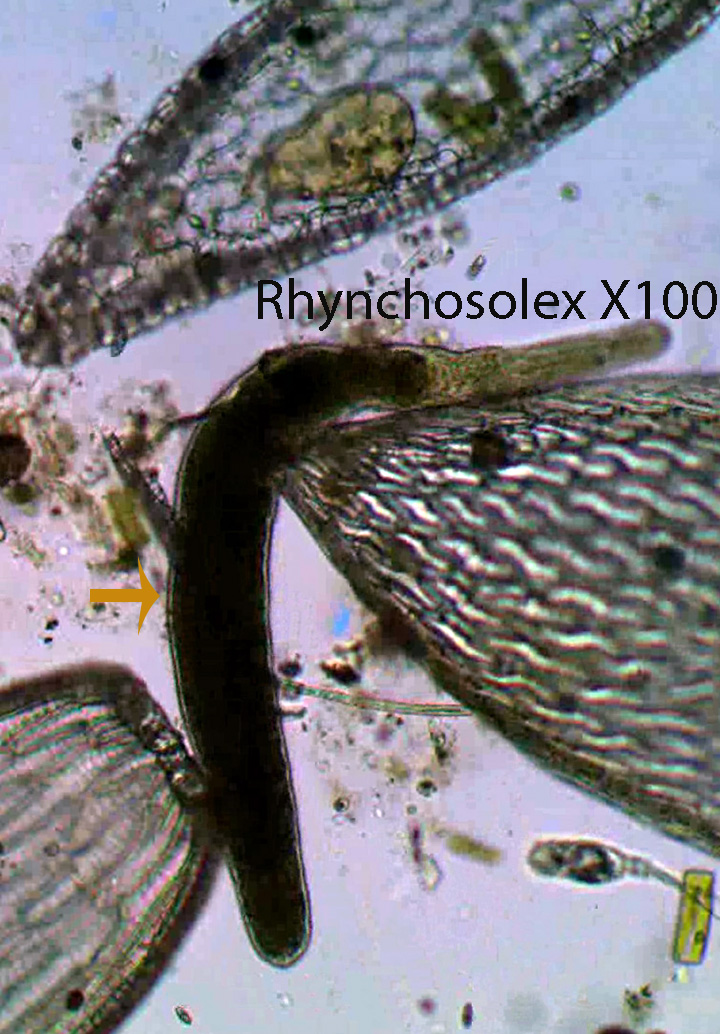
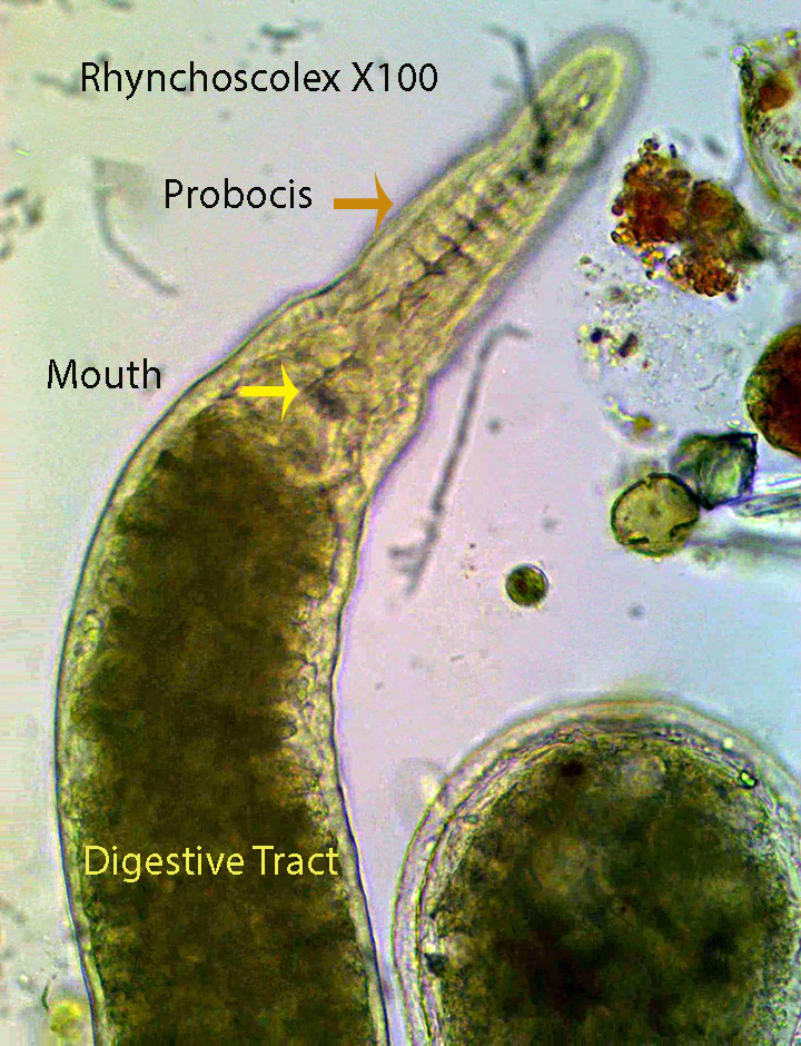
The anterior end of Rhynchoscolex ( about 1 mm long) terminates as a narrow “proboscis”. It reproduces asexually by dividing the body into a series of zooids that eventually break free and develop into adult worms. During active feeding bouts they may twist and turn using mesoderm derived, circular and longitudinal muscles.
Stenostomum (Stenostomidae)
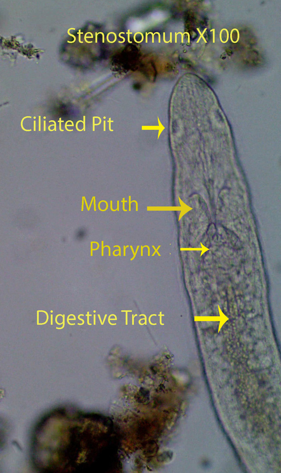
Stenostomum, about 1 mm long, is a small free-living flatworm that is common in freshwater environments. The animal glides smoothly through the water , propelled by ventral cilia. The flatworm lacks eyes and reproduces asexually by producing zooids. During active feeding bouts they may twist and turn using mesoderm derived, circular and longitudinal muscles. They have a digestive tract with only one opening that serves as both mouth and anus. One ciliated pit (Groove) is located, laterally, on each side of the head. The pits are visible toward the end of the video. The large mass inside the head is the brain. The mouth, just behind the brain, is connected to a short, muscular pharynx that leads into a sac-like digestive tract.
Gastrotrichs
Gastrotrichs are microscopic animals that are abundant in marine and freshwater habitats. They characteristically are flexible and flattened ventrally. Tracts of cilia on the ventral surface are responsible for their ability to glide smoothly over the substratum. Internal circular and longitudinal muscle contractions allow turning and twisting movements. The head is slightly rounded and bears lateral sensory cilia and a terminal mouth. Internally the mouth connects to a muscular pharynx that in turn is attached to a simple, tubular digestive tract that opens to the outside through the anus. The posterior end bears two pointed “toes” (Caudal Furcae) with internal adhesive glands that secrete a substance that can temporarily hold the animal in place during feeding bouts.
Chaetonotus (Chaetonotidae)
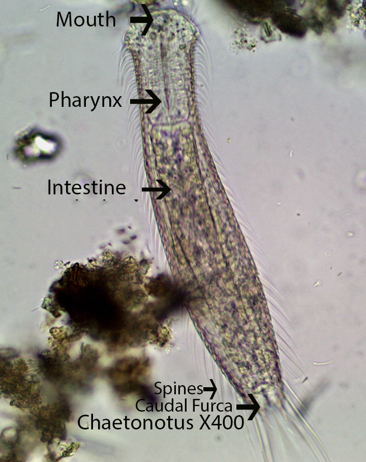
Chaetonotus,about 150 microns long, is covered with a cuticle that bears spines. They feed on algae, bacteria, protozoans and detritus.
Ichthydium (Chaetonotidae)

Ichthydium, about 150 microns long, has a smooth cuticle. They feed on algae, bacteria, protozoans and detritus.

Leave a comment